Calcium »
PDB 2tga-2v5c »
2v02 »
Calcium in PDB 2v02: Recombinant Vertebrate Calmodulin Complexed with Ba
Protein crystallography data
The structure of Recombinant Vertebrate Calmodulin Complexed with Ba, PDB code: 2v02
was solved by
P.Kursula,
V.Majava,
with X-Ray Crystallography technique. A brief refinement statistics is given in the table below:
| Resolution Low / High (Å) | 20.00 / 2.20 |
| Space group | P 1 |
| Cell size a, b, c (Å), α, β, γ (°) | 24.410, 29.320, 53.320, 89.84, 85.68, 82.80 |
| R / Rfree (%) | 24.3 / 29.4 |
Other elements in 2v02:
The structure of Recombinant Vertebrate Calmodulin Complexed with Ba also contains other interesting chemical elements:
| Barium | (Ba) | 2 atoms |
Calcium Binding Sites:
The binding sites of Calcium atom in the Recombinant Vertebrate Calmodulin Complexed with Ba
(pdb code 2v02). This binding sites where shown within
5.0 Angstroms radius around Calcium atom.
In total 3 binding sites of Calcium where determined in the Recombinant Vertebrate Calmodulin Complexed with Ba, PDB code: 2v02:
Jump to Calcium binding site number: 1; 2; 3;
In total 3 binding sites of Calcium where determined in the Recombinant Vertebrate Calmodulin Complexed with Ba, PDB code: 2v02:
Jump to Calcium binding site number: 1; 2; 3;
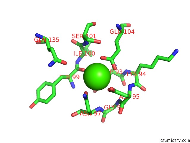
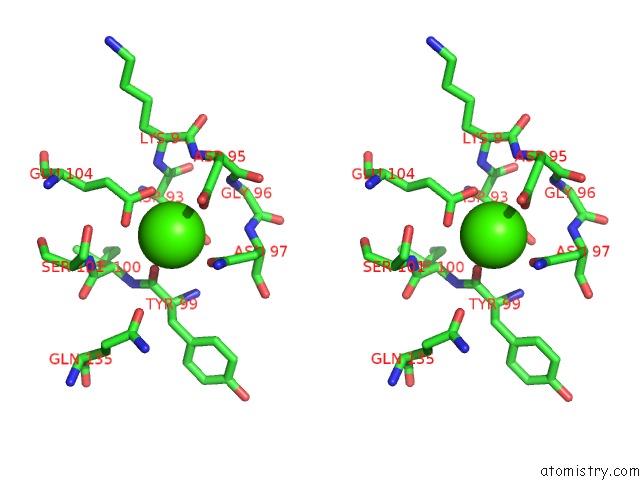
Calcium binding site 1 out of 3 in 2v02
Go back to
Calcium binding site 1 out
of 3 in the Recombinant Vertebrate Calmodulin Complexed with Ba
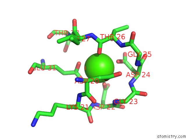
Mono view
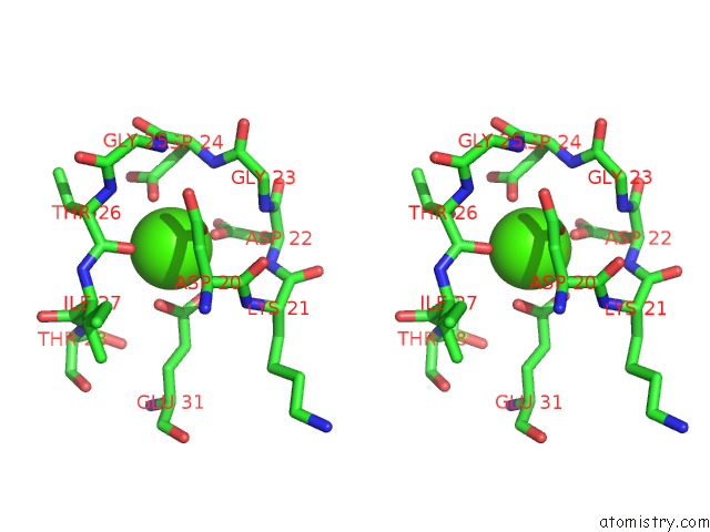
Stereo pair view
Mono view
Stereo pair view
A full contact list of Calcium with other atoms in the Ca binding
site number 1 of Recombinant Vertebrate Calmodulin Complexed with Ba within 5.0Å range:
|
Calcium binding site 2 out of 3 in 2v02
Go back to
Calcium binding site 2 out
of 3 in the Recombinant Vertebrate Calmodulin Complexed with Ba
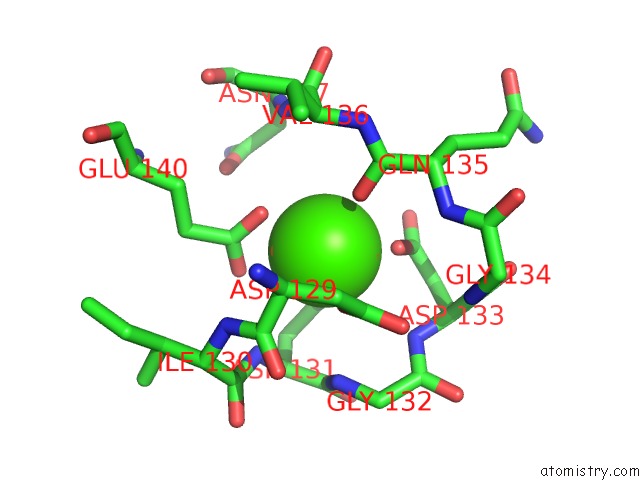
Mono view
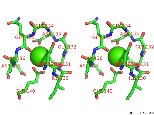
Stereo pair view
Mono view
Stereo pair view
A full contact list of Calcium with other atoms in the Ca binding
site number 2 of Recombinant Vertebrate Calmodulin Complexed with Ba within 5.0Å range:
|
Calcium binding site 3 out of 3 in 2v02
Go back to
Calcium binding site 3 out
of 3 in the Recombinant Vertebrate Calmodulin Complexed with Ba
Mono view
Stereo pair view
Mono view
Stereo pair view
A full contact list of Calcium with other atoms in the Ca binding
site number 3 of Recombinant Vertebrate Calmodulin Complexed with Ba within 5.0Å range:
|
Reference:
P.Kursula,
V.Majava.
A Structural Insight Into Lead Neurotoxicity and Calmodulin Activation By Heavy Metals. Acta Crystallogr.,Sect.F V. 63 653 2007.
ISSN: ESSN 1744-3091
PubMed: 17671360
DOI: 10.1107/S1744309107034525
Page generated: Fri Jul 12 17:48:19 2024
ISSN: ESSN 1744-3091
PubMed: 17671360
DOI: 10.1107/S1744309107034525
Last articles
Zn in 9MJ5Zn in 9HNW
Zn in 9G0L
Zn in 9FNE
Zn in 9DZN
Zn in 9E0I
Zn in 9D32
Zn in 9DAK
Zn in 8ZXC
Zn in 8ZUF