Calcium »
PDB 2tga-2v5c »
2v51 »
Calcium in PDB 2v51: Structure of Mal-RPEL1 Complexed to Actin
Protein crystallography data
The structure of Structure of Mal-RPEL1 Complexed to Actin, PDB code: 2v51
was solved by
S.Mouilleron,
S.Guettler,
C.A.Langer,
R.Treisman,
N.Q.Mcdonald,
with X-Ray Crystallography technique. A brief refinement statistics is given in the table below:
| Resolution Low / High (Å) | 20.00 / 2.35 |
| Space group | P 1 21 1 |
| Cell size a, b, c (Å), α, β, γ (°) | 53.092, 59.071, 166.203, 90.00, 90.06, 90.00 |
| R / Rfree (%) | 18.3 / 24.8 |
Calcium Binding Sites:
The binding sites of Calcium atom in the Structure of Mal-RPEL1 Complexed to Actin
(pdb code 2v51). This binding sites where shown within
5.0 Angstroms radius around Calcium atom.
In total 2 binding sites of Calcium where determined in the Structure of Mal-RPEL1 Complexed to Actin, PDB code: 2v51:
Jump to Calcium binding site number: 1; 2;
In total 2 binding sites of Calcium where determined in the Structure of Mal-RPEL1 Complexed to Actin, PDB code: 2v51:
Jump to Calcium binding site number: 1; 2;
Calcium binding site 1 out of 2 in 2v51
Go back to
Calcium binding site 1 out
of 2 in the Structure of Mal-RPEL1 Complexed to Actin
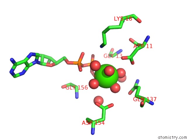
Mono view
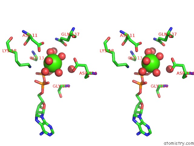
Stereo pair view
Mono view
Stereo pair view
A full contact list of Calcium with other atoms in the Ca binding
site number 1 of Structure of Mal-RPEL1 Complexed to Actin within 5.0Å range:
|
Calcium binding site 2 out of 2 in 2v51
Go back to
Calcium binding site 2 out
of 2 in the Structure of Mal-RPEL1 Complexed to Actin
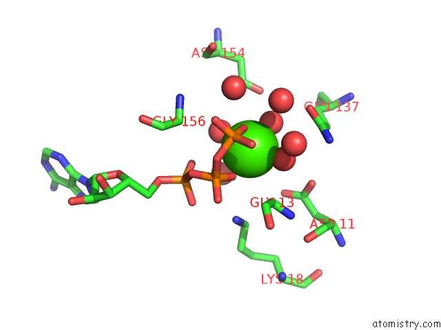
Mono view
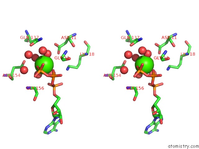
Stereo pair view
Mono view
Stereo pair view
A full contact list of Calcium with other atoms in the Ca binding
site number 2 of Structure of Mal-RPEL1 Complexed to Actin within 5.0Å range:
|
Reference:
S.Mouilleron,
S.Guettler,
C.A.Langer,
R.Treisman,
N.Q.Mcdonald.
Molecular Basis For G-Actin Binding to Rpel Motifs From the Serum Response Factor Coactivator Mal. Embo J. V. 27 3198 2008.
ISSN: ESSN 1460-2075
PubMed: 19008859
DOI: 10.1038/EMBOJ.2008.235
Page generated: Fri Jul 12 17:49:48 2024
ISSN: ESSN 1460-2075
PubMed: 19008859
DOI: 10.1038/EMBOJ.2008.235
Last articles
Zn in 9J0NZn in 9J0O
Zn in 9J0P
Zn in 9FJX
Zn in 9EKB
Zn in 9C0F
Zn in 9CAH
Zn in 9CH0
Zn in 9CH3
Zn in 9CH1