Calcium »
PDB 2v5d-2vjj »
2v9m »
Calcium in PDB 2v9m: L-Rhamnulose-1-Phosphate Aldolase From Escherichia Coli (Mutant A87M- T109F-E192A)
Enzymatic activity of L-Rhamnulose-1-Phosphate Aldolase From Escherichia Coli (Mutant A87M- T109F-E192A)
All present enzymatic activity of L-Rhamnulose-1-Phosphate Aldolase From Escherichia Coli (Mutant A87M- T109F-E192A):
4.1.2.19;
4.1.2.19;
Protein crystallography data
The structure of L-Rhamnulose-1-Phosphate Aldolase From Escherichia Coli (Mutant A87M- T109F-E192A), PDB code: 2v9m
was solved by
D.Grueninger,
G.E.Schulz,
with X-Ray Crystallography technique. A brief refinement statistics is given in the table below:
| Resolution Low / High (Å) | 20.00 / 1.30 |
| Space group | P 4 |
| Cell size a, b, c (Å), α, β, γ (°) | 83.407, 83.407, 97.299, 90.00, 90.00, 90.00 |
| R / Rfree (%) | 10.4 / 14.1 |
Other elements in 2v9m:
The structure of L-Rhamnulose-1-Phosphate Aldolase From Escherichia Coli (Mutant A87M- T109F-E192A) also contains other interesting chemical elements:
| Zinc | (Zn) | 4 atoms |
Calcium Binding Sites:
The binding sites of Calcium atom in the L-Rhamnulose-1-Phosphate Aldolase From Escherichia Coli (Mutant A87M- T109F-E192A)
(pdb code 2v9m). This binding sites where shown within
5.0 Angstroms radius around Calcium atom.
In total only one binding site of Calcium was determined in the L-Rhamnulose-1-Phosphate Aldolase From Escherichia Coli (Mutant A87M- T109F-E192A), PDB code: 2v9m:
In total only one binding site of Calcium was determined in the L-Rhamnulose-1-Phosphate Aldolase From Escherichia Coli (Mutant A87M- T109F-E192A), PDB code: 2v9m:
Calcium binding site 1 out of 1 in 2v9m
Go back to
Calcium binding site 1 out
of 1 in the L-Rhamnulose-1-Phosphate Aldolase From Escherichia Coli (Mutant A87M- T109F-E192A)
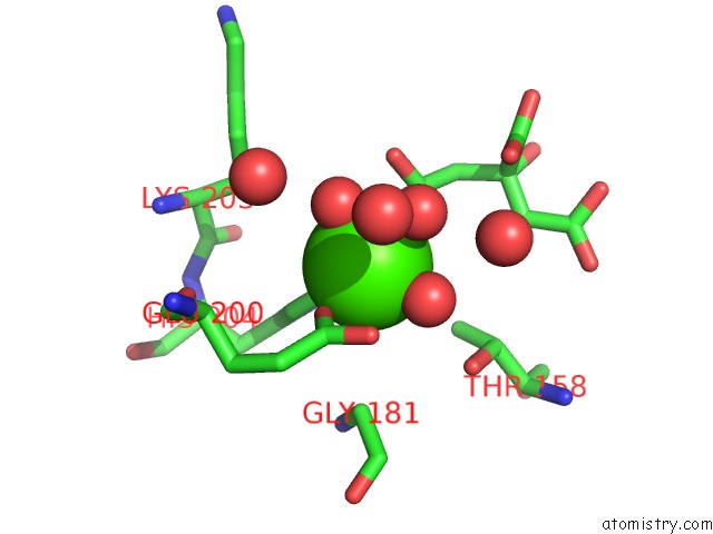
Mono view
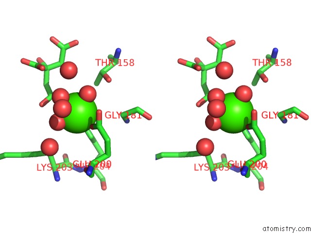
Stereo pair view
Mono view
Stereo pair view
A full contact list of Calcium with other atoms in the Ca binding
site number 1 of L-Rhamnulose-1-Phosphate Aldolase From Escherichia Coli (Mutant A87M- T109F-E192A) within 5.0Å range:
|
Reference:
D.Grueninger,
N.Treiber,
M.O.P.Ziegler,
J.W.A.Koetter,
M.-S.Schulze,
G.E.Schulz.
Designed Protein-Protein Association. Science V. 319 206 2008.
ISSN: ISSN 0036-8075
PubMed: 18187656
DOI: 10.1126/SCIENCE.1150421
Page generated: Tue Jul 8 08:30:54 2025
ISSN: ISSN 0036-8075
PubMed: 18187656
DOI: 10.1126/SCIENCE.1150421
Last articles
Cl in 5RAJCl in 5RAM
Cl in 5RAL
Cl in 5RAK
Cl in 5RAF
Cl in 5RAG
Cl in 5RAH
Cl in 5RAI
Cl in 5RAE
Cl in 5RAD