Calcium »
PDB 2v5d-2vjj »
2vf9 »
Calcium in PDB 2vf9: Crystal Structure of Bacteriophage PRR1
Protein crystallography data
The structure of Crystal Structure of Bacteriophage PRR1, PDB code: 2vf9
was solved by
M.Persson,
K.Tars,
L.Liljas,
with X-Ray Crystallography technique. A brief refinement statistics is given in the table below:
| Resolution Low / High (Å) | 29.12 / 3.50 |
| Space group | P 1 |
| Cell size a, b, c (Å), α, β, γ (°) | 281.400, 285.400, 472.500, 93.27, 90.00, 119.44 |
| R / Rfree (%) | 31.1 / 31.3 |
Calcium Binding Sites:
The binding sites of Calcium atom in the Crystal Structure of Bacteriophage PRR1
(pdb code 2vf9). This binding sites where shown within
5.0 Angstroms radius around Calcium atom.
In total only one binding site of Calcium was determined in the Crystal Structure of Bacteriophage PRR1, PDB code: 2vf9:
In total only one binding site of Calcium was determined in the Crystal Structure of Bacteriophage PRR1, PDB code: 2vf9:
Calcium binding site 1 out of 1 in 2vf9
Go back to
Calcium binding site 1 out
of 1 in the Crystal Structure of Bacteriophage PRR1

Mono view
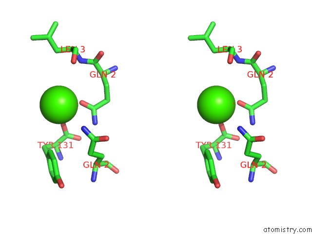
Stereo pair view
Mono view
Stereo pair view
A full contact list of Calcium with other atoms in the Ca binding
site number 1 of Crystal Structure of Bacteriophage PRR1 within 5.0Å range:
|
Reference:
M.Persson,
K.Tars,
L.Liljas.
The Capsid of the Small Rna Phage PRR1 Is Stabilized By Metal Ions J.Mol.Biol. V. 383 914 2008.
ISSN: ISSN 0022-2836
PubMed: 18786545
DOI: 10.1016/J.JMB.2008.08.060
Page generated: Fri Jul 12 18:01:09 2024
ISSN: ISSN 0022-2836
PubMed: 18786545
DOI: 10.1016/J.JMB.2008.08.060
Last articles
Zn in 9J0NZn in 9J0O
Zn in 9J0P
Zn in 9FJX
Zn in 9EKB
Zn in 9C0F
Zn in 9CAH
Zn in 9CH0
Zn in 9CH3
Zn in 9CH1