Calcium »
PDB 2vk5-2vuz »
2vmc »
Calcium in PDB 2vmc: Structure of the Complex of Discoidin II From Dictyostelium Discoideum with N-Acetyl-Galactosamine
Protein crystallography data
The structure of Structure of the Complex of Discoidin II From Dictyostelium Discoideum with N-Acetyl-Galactosamine, PDB code: 2vmc
was solved by
K.Saboia,
M.Satre,
A.Imberty,
A.Varrot,
with X-Ray Crystallography technique. A brief refinement statistics is given in the table below:
| Resolution Low / High (Å) | 43.60 / 1.90 |
| Space group | H 3 2 |
| Cell size a, b, c (Å), α, β, γ (°) | 82.520, 82.520, 261.600, 90.00, 90.00, 120.00 |
| R / Rfree (%) | 15.7 / 19.8 |
Other elements in 2vmc:
The structure of Structure of the Complex of Discoidin II From Dictyostelium Discoideum with N-Acetyl-Galactosamine also contains other interesting chemical elements:
| Chlorine | (Cl) | 2 atoms |
Calcium Binding Sites:
The binding sites of Calcium atom in the Structure of the Complex of Discoidin II From Dictyostelium Discoideum with N-Acetyl-Galactosamine
(pdb code 2vmc). This binding sites where shown within
5.0 Angstroms radius around Calcium atom.
In total only one binding site of Calcium was determined in the Structure of the Complex of Discoidin II From Dictyostelium Discoideum with N-Acetyl-Galactosamine, PDB code: 2vmc:
In total only one binding site of Calcium was determined in the Structure of the Complex of Discoidin II From Dictyostelium Discoideum with N-Acetyl-Galactosamine, PDB code: 2vmc:
Calcium binding site 1 out of 1 in 2vmc
Go back to
Calcium binding site 1 out
of 1 in the Structure of the Complex of Discoidin II From Dictyostelium Discoideum with N-Acetyl-Galactosamine
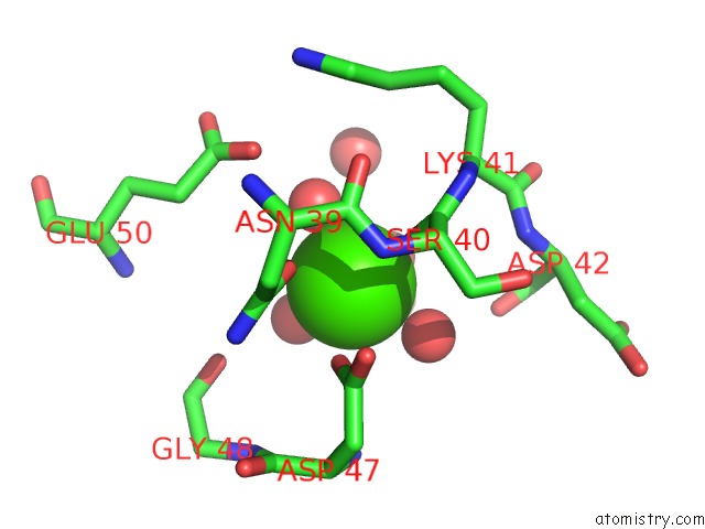
Mono view
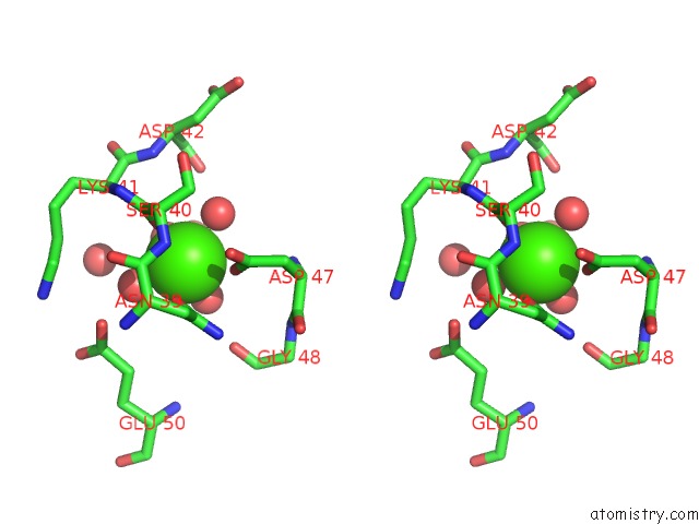
Stereo pair view
Mono view
Stereo pair view
A full contact list of Calcium with other atoms in the Ca binding
site number 1 of Structure of the Complex of Discoidin II From Dictyostelium Discoideum with N-Acetyl-Galactosamine within 5.0Å range:
|
Reference:
K.S.Aragao,
M.Satre,
A.Imberty,
A.Varrot.
Structure Determination of Discoidin II From Dictyostelium Discoideum and Carbohydrate Binding Properties of the Lectin Domain. Proteins V. 73 43 2008.
ISSN: ISSN 0887-3585
PubMed: 18384150
DOI: 10.1002/PROT.22038
Page generated: Fri Jul 12 18:04:39 2024
ISSN: ISSN 0887-3585
PubMed: 18384150
DOI: 10.1002/PROT.22038
Last articles
Zn in 9JYWZn in 9IR4
Zn in 9IR3
Zn in 9GMX
Zn in 9GMW
Zn in 9JEJ
Zn in 9ERF
Zn in 9ERE
Zn in 9EGV
Zn in 9EGW