Calcium »
PDB 2vk5-2vuz »
2vs7 »
Calcium in PDB 2vs7: The Crystal Structure of I-Dmoi in Complex with Dna and Ca
Protein crystallography data
The structure of The Crystal Structure of I-Dmoi in Complex with Dna and Ca, PDB code: 2vs7
was solved by
M.J.Marcaida,
J.Prieto,
P.Redondo,
A.D.Nadra,
A.Alibes,
L.Serrano,
S.Grizot,
P.Duchateau,
F.Paques,
F.J.Blanco,
G.Montoya,
with X-Ray Crystallography technique. A brief refinement statistics is given in the table below:
| Resolution Low / High (Å) | 25.76 / 2.05 |
| Space group | P 1 21 1 |
| Cell size a, b, c (Å), α, β, γ (°) | 106.988, 70.587, 106.848, 90.00, 119.75, 90.00 |
| R / Rfree (%) | 18.7 / 22.8 |
Calcium Binding Sites:
The binding sites of Calcium atom in the The Crystal Structure of I-Dmoi in Complex with Dna and Ca
(pdb code 2vs7). This binding sites where shown within
5.0 Angstroms radius around Calcium atom.
In total 3 binding sites of Calcium where determined in the The Crystal Structure of I-Dmoi in Complex with Dna and Ca, PDB code: 2vs7:
Jump to Calcium binding site number: 1; 2; 3;
In total 3 binding sites of Calcium where determined in the The Crystal Structure of I-Dmoi in Complex with Dna and Ca, PDB code: 2vs7:
Jump to Calcium binding site number: 1; 2; 3;
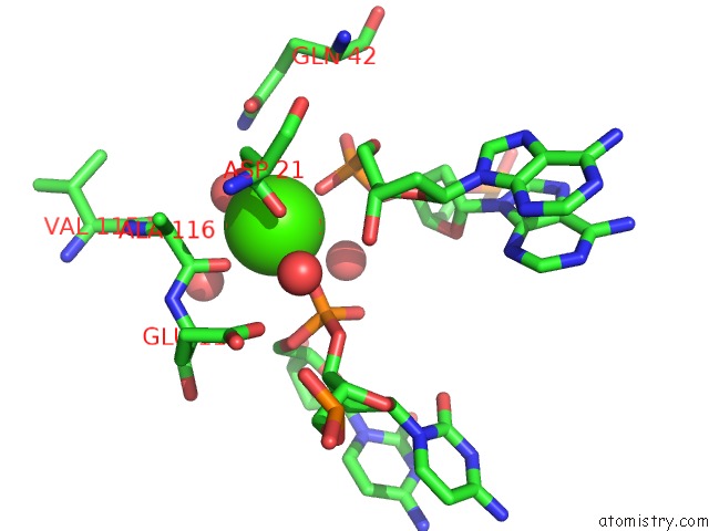
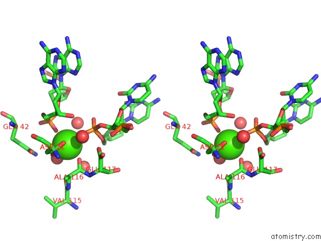
Calcium binding site 1 out of 3 in 2vs7
Go back to
Calcium binding site 1 out
of 3 in the The Crystal Structure of I-Dmoi in Complex with Dna and Ca
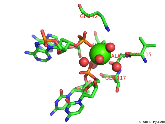
Mono view
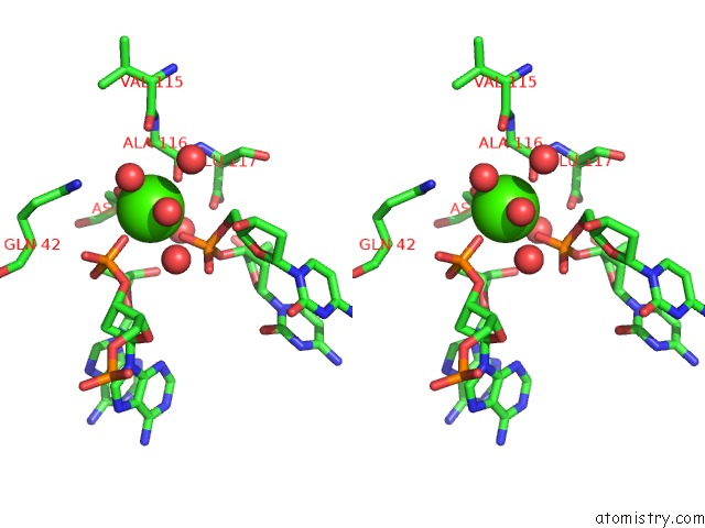
Stereo pair view
Mono view
Stereo pair view
A full contact list of Calcium with other atoms in the Ca binding
site number 1 of The Crystal Structure of I-Dmoi in Complex with Dna and Ca within 5.0Å range:
|
Calcium binding site 2 out of 3 in 2vs7
Go back to
Calcium binding site 2 out
of 3 in the The Crystal Structure of I-Dmoi in Complex with Dna and Ca
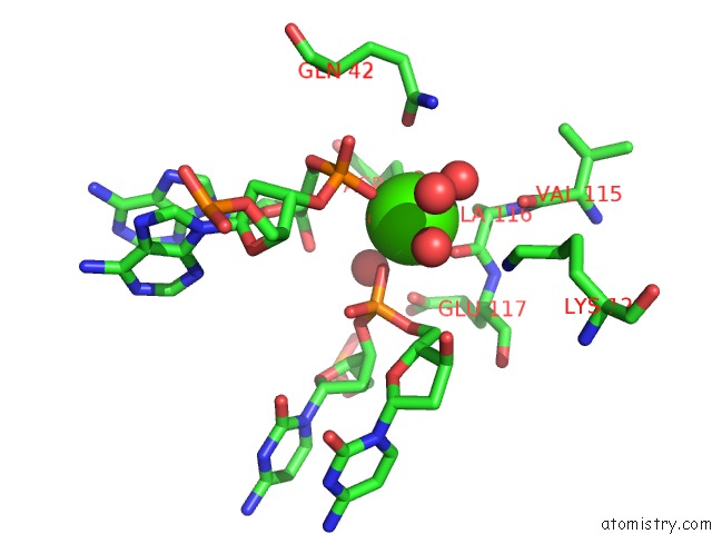
Mono view
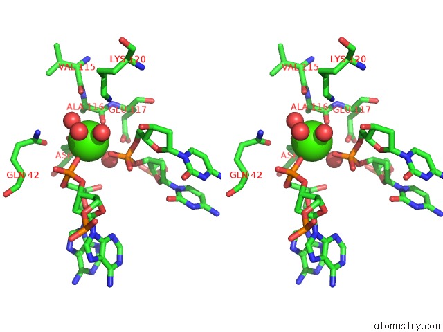
Stereo pair view
Mono view
Stereo pair view
A full contact list of Calcium with other atoms in the Ca binding
site number 2 of The Crystal Structure of I-Dmoi in Complex with Dna and Ca within 5.0Å range:
|
Calcium binding site 3 out of 3 in 2vs7
Go back to
Calcium binding site 3 out
of 3 in the The Crystal Structure of I-Dmoi in Complex with Dna and Ca
Mono view
Stereo pair view
Mono view
Stereo pair view
A full contact list of Calcium with other atoms in the Ca binding
site number 3 of The Crystal Structure of I-Dmoi in Complex with Dna and Ca within 5.0Å range:
|
Reference:
M.J.Marcaida,
J.Prieto,
P.Redondo,
A.D.Nadra,
A.Alibes,
L.Serrano,
S.Grizot,
P.Duchateau,
F.Paques,
F.J.Blanco,
G.Montoya.
Crystal Structure of I-Dmoi in Complex with Its Target Dna Provides New Insights Into Meganuclease Engineering. Proc.Nat.Acad.Sci.Usa V. 105 16888 2008.
ISSN: ISSN 0027-8424
PubMed: 18974222
DOI: 10.1073/PNAS.0804795105
Page generated: Tue Jul 8 08:43:59 2025
ISSN: ISSN 0027-8424
PubMed: 18974222
DOI: 10.1073/PNAS.0804795105
Last articles
Cl in 5HJACl in 5HJC
Cl in 5HI5
Cl in 5HI4
Cl in 5HI2
Cl in 5HH4
Cl in 5HGL
Cl in 5HGK
Cl in 5HGI
Cl in 5HG1