Calcium »
PDB 2vvc-2w3j »
2vy0 »
Calcium in PDB 2vy0: The X-Ray Structure of Endo-Beta-1,3-Glucanase From Pyrococcus Furiosus
Enzymatic activity of The X-Ray Structure of Endo-Beta-1,3-Glucanase From Pyrococcus Furiosus
All present enzymatic activity of The X-Ray Structure of Endo-Beta-1,3-Glucanase From Pyrococcus Furiosus:
3.2.1.39;
3.2.1.39;
Protein crystallography data
The structure of The X-Ray Structure of Endo-Beta-1,3-Glucanase From Pyrococcus Furiosus, PDB code: 2vy0
was solved by
A.Ilari,
A.Fiorillo,
with X-Ray Crystallography technique. A brief refinement statistics is given in the table below:
| Resolution Low / High (Å) | 50.00 / 2.16 |
| Space group | P 1 21 1 |
| Cell size a, b, c (Å), α, β, γ (°) | 44.363, 84.762, 69.232, 90.00, 104.97, 90.00 |
| R / Rfree (%) | 19.1 / 22.9 |
Other elements in 2vy0:
The structure of The X-Ray Structure of Endo-Beta-1,3-Glucanase From Pyrococcus Furiosus also contains other interesting chemical elements:
| Chlorine | (Cl) | 3 atoms |
| Sodium | (Na) | 3 atoms |
Calcium Binding Sites:
The binding sites of Calcium atom in the The X-Ray Structure of Endo-Beta-1,3-Glucanase From Pyrococcus Furiosus
(pdb code 2vy0). This binding sites where shown within
5.0 Angstroms radius around Calcium atom.
In total 2 binding sites of Calcium where determined in the The X-Ray Structure of Endo-Beta-1,3-Glucanase From Pyrococcus Furiosus, PDB code: 2vy0:
Jump to Calcium binding site number: 1; 2;
In total 2 binding sites of Calcium where determined in the The X-Ray Structure of Endo-Beta-1,3-Glucanase From Pyrococcus Furiosus, PDB code: 2vy0:
Jump to Calcium binding site number: 1; 2;
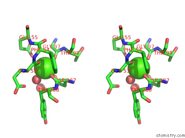
Calcium binding site 1 out of 2 in 2vy0
Go back to
Calcium binding site 1 out
of 2 in the The X-Ray Structure of Endo-Beta-1,3-Glucanase From Pyrococcus Furiosus
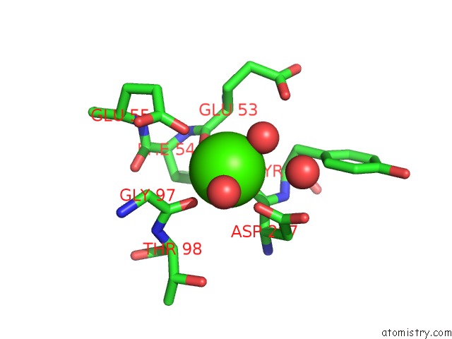
Mono view

Stereo pair view
Mono view
Stereo pair view
A full contact list of Calcium with other atoms in the Ca binding
site number 1 of The X-Ray Structure of Endo-Beta-1,3-Glucanase From Pyrococcus Furiosus within 5.0Å range:
|
Calcium binding site 2 out of 2 in 2vy0
Go back to
Calcium binding site 2 out
of 2 in the The X-Ray Structure of Endo-Beta-1,3-Glucanase From Pyrococcus Furiosus
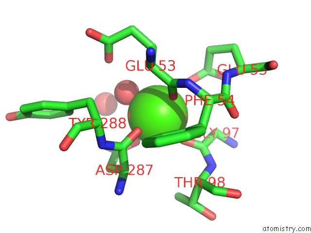
Mono view
Stereo pair view
Mono view
Stereo pair view
A full contact list of Calcium with other atoms in the Ca binding
site number 2 of The X-Ray Structure of Endo-Beta-1,3-Glucanase From Pyrococcus Furiosus within 5.0Å range:
|
Reference:
A.Ilari,
A.Fiorillo,
S.Angelaccio,
R.Florio,
R.Chiaraluce,
J.Van Der Oost,
V.Consalvi.
Crystal Structure of A Family 16 Endoglucanase From the Hyperthermophile Pyrococcus Furiosus- Structural Basis of Substrate Recognition. Febs J. V. 276 1048 2009.
ISSN: ISSN 1742-464X
PubMed: 19154353
DOI: 10.1111/J.1742-4658.2008.06848.X
Page generated: Fri Jul 12 18:16:06 2024
ISSN: ISSN 1742-464X
PubMed: 19154353
DOI: 10.1111/J.1742-4658.2008.06848.X
Last articles
Zn in 9J0NZn in 9J0O
Zn in 9J0P
Zn in 9FJX
Zn in 9EKB
Zn in 9C0F
Zn in 9CAH
Zn in 9CH0
Zn in 9CH3
Zn in 9CH1