Calcium »
PDB 2vvc-2w3j »
2w0c »
Calcium in PDB 2w0c: X-Ray Structure of the Entire Lipid-Containing Bacteriophage PM2
Protein crystallography data
The structure of X-Ray Structure of the Entire Lipid-Containing Bacteriophage PM2, PDB code: 2w0c
was solved by
N.G.A.Abrescia,
J.M.Grimes,
H.M.Kivela,
R.Assenberg,
G.C.Sutton,
S.J.Butcher,
J.K.H.Bamford,
D.H.Bamford,
D.I.Stuart,
with X-Ray Crystallography technique. A brief refinement statistics is given in the table below:
| Resolution Low / High (Å) | 96.00 / 7.00 |
| Space group | C 1 2 1 24 |
| Cell size a, b, c (Å), α, β, γ (°) | 946.900, 677.600, 1067.600, 90.00, 102.90, 90.00 |
| R / Rfree (%) | 41.9 / n/a |
Calcium Binding Sites:
Pages:
>>> Page 1 <<< Page 2, Binding sites: 11 - 11;Binding sites:
The binding sites of Calcium atom in the X-Ray Structure of the Entire Lipid-Containing Bacteriophage PM2 (pdb code 2w0c). This binding sites where shown within 5.0 Angstroms radius around Calcium atom.In total 11 binding sites of Calcium where determined in the X-Ray Structure of the Entire Lipid-Containing Bacteriophage PM2, PDB code: 2w0c:
Jump to Calcium binding site number: 1; 2; 3; 4; 5; 6; 7; 8; 9; 10;
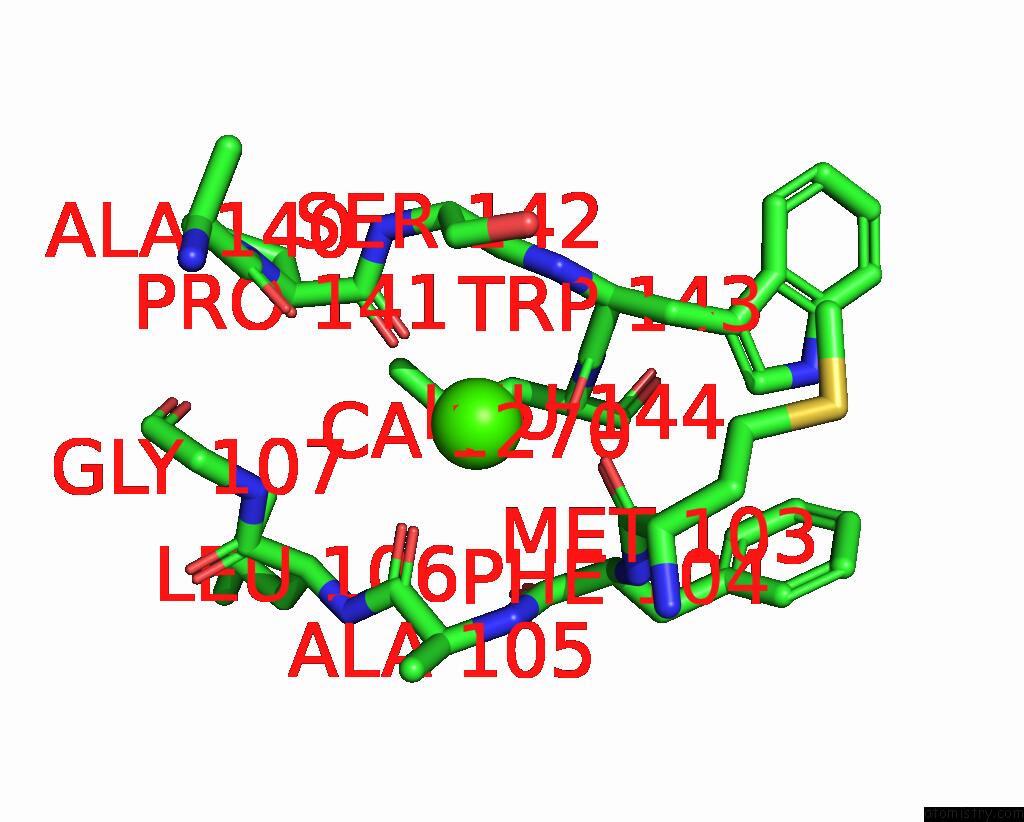
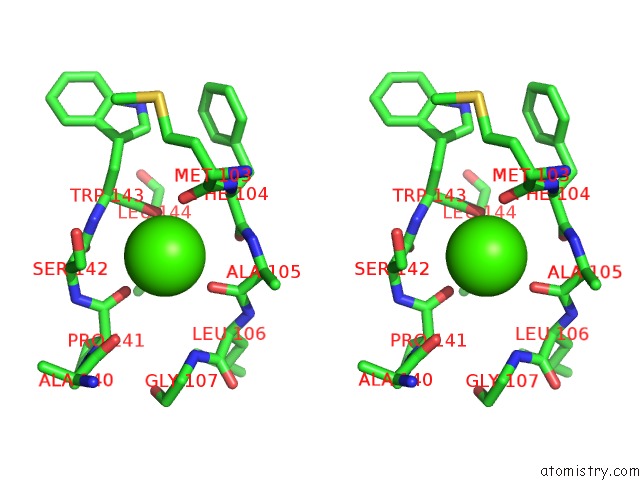
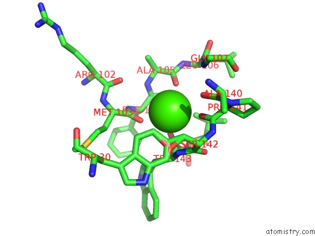
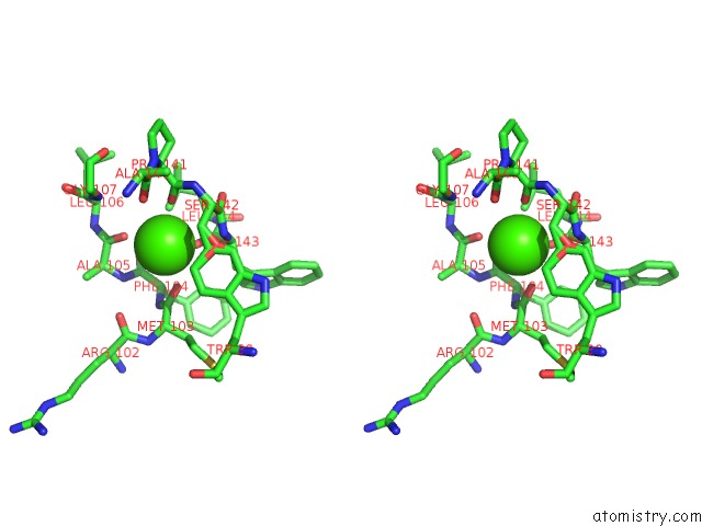
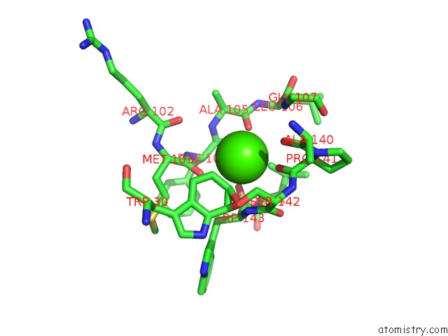
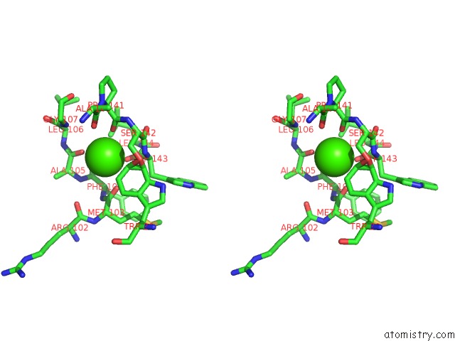
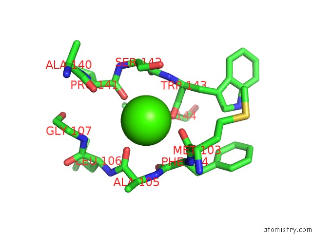
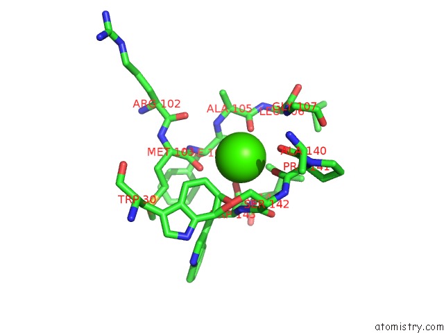
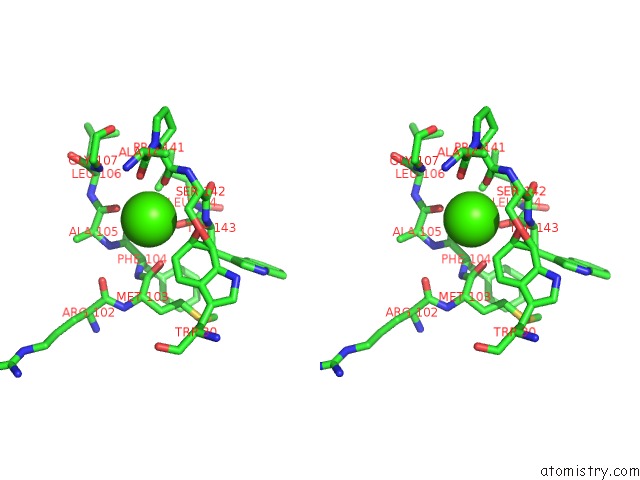
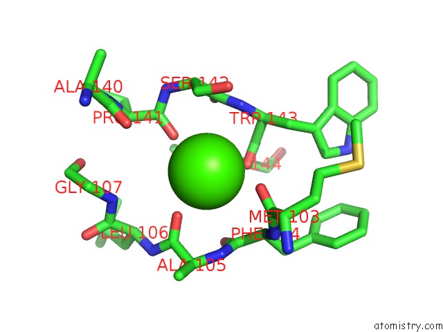
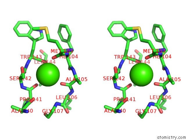
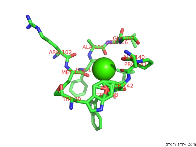
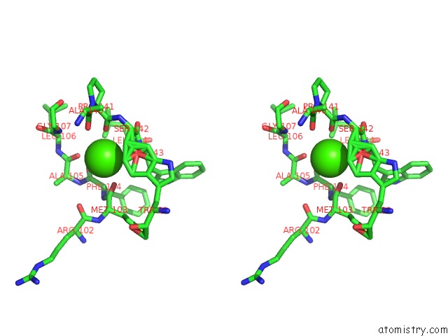
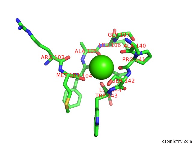
Calcium binding site 1 out of 11 in 2w0c
Go back to
Calcium binding site 1 out
of 11 in the X-Ray Structure of the Entire Lipid-Containing Bacteriophage PM2
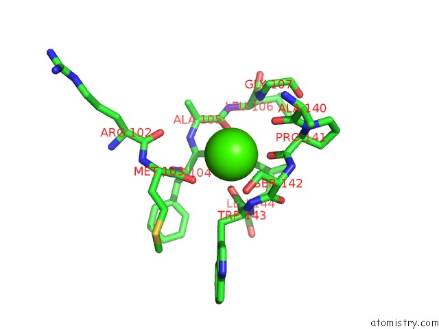
Mono view
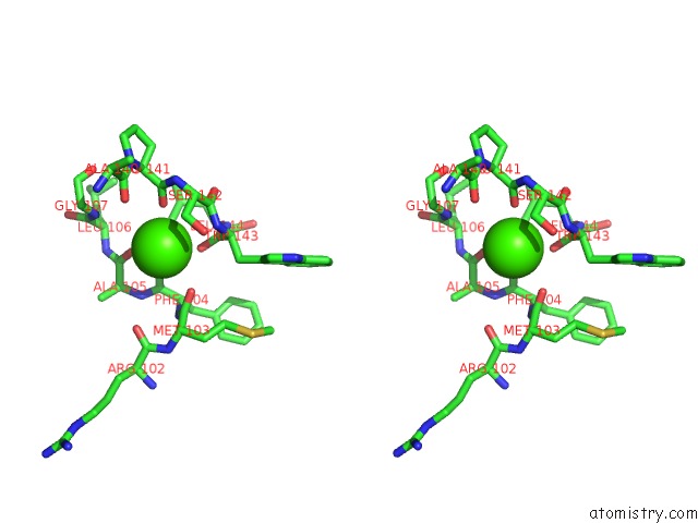
Stereo pair view
Mono view
Stereo pair view
A full contact list of Calcium with other atoms in the Ca binding
site number 1 of X-Ray Structure of the Entire Lipid-Containing Bacteriophage PM2 within 5.0Å range:
|
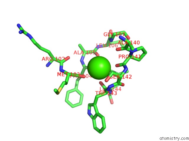
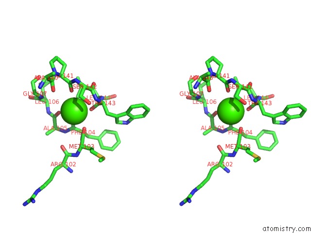
Calcium binding site 2 out of 11 in 2w0c
Go back to
Calcium binding site 2 out
of 11 in the X-Ray Structure of the Entire Lipid-Containing Bacteriophage PM2
Mono view
Stereo pair view
Mono view
Stereo pair view
A full contact list of Calcium with other atoms in the Ca binding
site number 2 of X-Ray Structure of the Entire Lipid-Containing Bacteriophage PM2 within 5.0Å range:
|
Calcium binding site 3 out of 11 in 2w0c
Go back to
Calcium binding site 3 out
of 11 in the X-Ray Structure of the Entire Lipid-Containing Bacteriophage PM2
Mono view
Stereo pair view
Mono view
Stereo pair view
A full contact list of Calcium with other atoms in the Ca binding
site number 3 of X-Ray Structure of the Entire Lipid-Containing Bacteriophage PM2 within 5.0Å range:
|
Calcium binding site 4 out of 11 in 2w0c
Go back to
Calcium binding site 4 out
of 11 in the X-Ray Structure of the Entire Lipid-Containing Bacteriophage PM2
Mono view
Stereo pair view
Mono view
Stereo pair view
A full contact list of Calcium with other atoms in the Ca binding
site number 4 of X-Ray Structure of the Entire Lipid-Containing Bacteriophage PM2 within 5.0Å range:
|
Calcium binding site 5 out of 11 in 2w0c
Go back to
Calcium binding site 5 out
of 11 in the X-Ray Structure of the Entire Lipid-Containing Bacteriophage PM2
Mono view
Stereo pair view
Mono view
Stereo pair view
A full contact list of Calcium with other atoms in the Ca binding
site number 5 of X-Ray Structure of the Entire Lipid-Containing Bacteriophage PM2 within 5.0Å range:
|
Calcium binding site 6 out of 11 in 2w0c
Go back to
Calcium binding site 6 out
of 11 in the X-Ray Structure of the Entire Lipid-Containing Bacteriophage PM2
Mono view

Stereo pair view
Mono view
Stereo pair view
A full contact list of Calcium with other atoms in the Ca binding
site number 6 of X-Ray Structure of the Entire Lipid-Containing Bacteriophage PM2 within 5.0Å range:
|
Calcium binding site 7 out of 11 in 2w0c
Go back to
Calcium binding site 7 out
of 11 in the X-Ray Structure of the Entire Lipid-Containing Bacteriophage PM2
Mono view
Stereo pair view
Mono view
Stereo pair view
A full contact list of Calcium with other atoms in the Ca binding
site number 7 of X-Ray Structure of the Entire Lipid-Containing Bacteriophage PM2 within 5.0Å range:
|
Calcium binding site 8 out of 11 in 2w0c
Go back to
Calcium binding site 8 out
of 11 in the X-Ray Structure of the Entire Lipid-Containing Bacteriophage PM2
Mono view
Stereo pair view
Mono view
Stereo pair view
A full contact list of Calcium with other atoms in the Ca binding
site number 8 of X-Ray Structure of the Entire Lipid-Containing Bacteriophage PM2 within 5.0Å range:
|
Calcium binding site 9 out of 11 in 2w0c
Go back to
Calcium binding site 9 out
of 11 in the X-Ray Structure of the Entire Lipid-Containing Bacteriophage PM2
Mono view
Stereo pair view
Mono view
Stereo pair view
A full contact list of Calcium with other atoms in the Ca binding
site number 9 of X-Ray Structure of the Entire Lipid-Containing Bacteriophage PM2 within 5.0Å range:
|
Calcium binding site 10 out of 11 in 2w0c
Go back to
Calcium binding site 10 out
of 11 in the X-Ray Structure of the Entire Lipid-Containing Bacteriophage PM2
Mono view

Stereo pair view
Mono view
Stereo pair view
A full contact list of Calcium with other atoms in the Ca binding
site number 10 of X-Ray Structure of the Entire Lipid-Containing Bacteriophage PM2 within 5.0Å range:
|
Reference:
N.G.A.Abrescia,
J.M.Grimes,
H.M.Kivela,
R.Assenberg,
G.C.Sutton,
S.J.Butcher,
J.K.H.Bamford,
D.H.Bamford,
D.I.Stuart.
Insights Into Virus Evolution and Membrane Biogenesis From the Structure of the Marine Lipid-Containing Bacteriophage PM2 Mol.Cell V. 31 749 2008.
ISSN: ISSN 1097-2765
PubMed: 18775333
DOI: 10.1016/J.MOLCEL.2008.06.026
Page generated: Fri Jul 12 18:18:45 2024
ISSN: ISSN 1097-2765
PubMed: 18775333
DOI: 10.1016/J.MOLCEL.2008.06.026
Last articles
Zn in 9J0NZn in 9J0O
Zn in 9J0P
Zn in 9FJX
Zn in 9EKB
Zn in 9C0F
Zn in 9CAH
Zn in 9CH0
Zn in 9CH3
Zn in 9CH1