Calcium »
PDB 2vvc-2w3j »
2w27 »
Calcium in PDB 2w27: Crystal Structure of the Bacillus Subtilis Ykui Protein, with An Eal Domain, in Complex with Substrate C-Di-Gmp and Calcium
Protein crystallography data
The structure of Crystal Structure of the Bacillus Subtilis Ykui Protein, with An Eal Domain, in Complex with Substrate C-Di-Gmp and Calcium, PDB code: 2w27
was solved by
S.Padavattan,
W.F.Anderson,
T.Schirmer,
with X-Ray Crystallography technique. A brief refinement statistics is given in the table below:
| Resolution Low / High (Å) | 30.00 / 2.80 |
| Space group | P 21 21 21 |
| Cell size a, b, c (Å), α, β, γ (°) | 46.167, 124.521, 168.751, 90.00, 90.00, 90.00 |
| R / Rfree (%) | 22.6 / 27.5 |
Calcium Binding Sites:
The binding sites of Calcium atom in the Crystal Structure of the Bacillus Subtilis Ykui Protein, with An Eal Domain, in Complex with Substrate C-Di-Gmp and Calcium
(pdb code 2w27). This binding sites where shown within
5.0 Angstroms radius around Calcium atom.
In total 2 binding sites of Calcium where determined in the Crystal Structure of the Bacillus Subtilis Ykui Protein, with An Eal Domain, in Complex with Substrate C-Di-Gmp and Calcium, PDB code: 2w27:
Jump to Calcium binding site number: 1; 2;
In total 2 binding sites of Calcium where determined in the Crystal Structure of the Bacillus Subtilis Ykui Protein, with An Eal Domain, in Complex with Substrate C-Di-Gmp and Calcium, PDB code: 2w27:
Jump to Calcium binding site number: 1; 2;
Calcium binding site 1 out of 2 in 2w27
Go back to
Calcium binding site 1 out
of 2 in the Crystal Structure of the Bacillus Subtilis Ykui Protein, with An Eal Domain, in Complex with Substrate C-Di-Gmp and Calcium
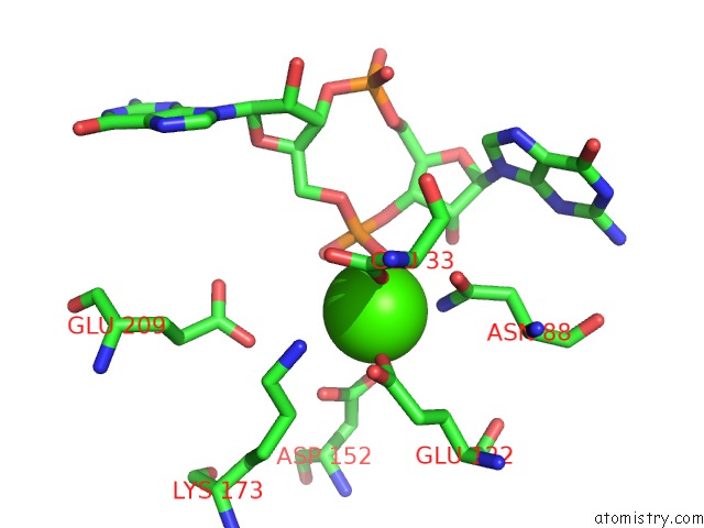
Mono view
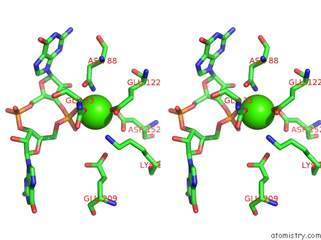
Stereo pair view
Mono view
Stereo pair view
A full contact list of Calcium with other atoms in the Ca binding
site number 1 of Crystal Structure of the Bacillus Subtilis Ykui Protein, with An Eal Domain, in Complex with Substrate C-Di-Gmp and Calcium within 5.0Å range:
|
Calcium binding site 2 out of 2 in 2w27
Go back to
Calcium binding site 2 out
of 2 in the Crystal Structure of the Bacillus Subtilis Ykui Protein, with An Eal Domain, in Complex with Substrate C-Di-Gmp and Calcium
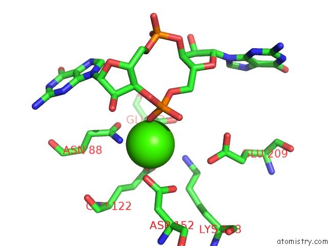
Mono view
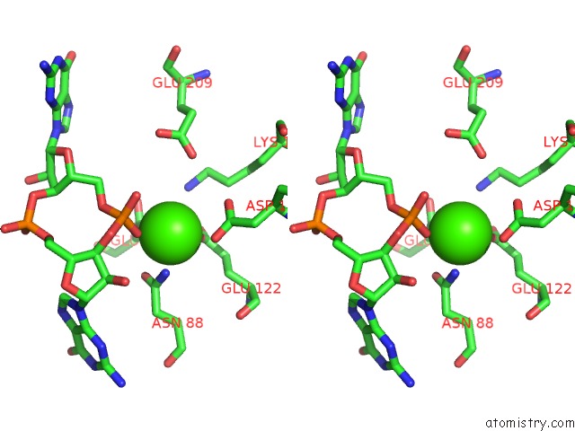
Stereo pair view
Mono view
Stereo pair view
A full contact list of Calcium with other atoms in the Ca binding
site number 2 of Crystal Structure of the Bacillus Subtilis Ykui Protein, with An Eal Domain, in Complex with Substrate C-Di-Gmp and Calcium within 5.0Å range:
|
Reference:
G.Minasov,
S.Padavattan,
L.Shuvalova,
J.S.Brunzelle,
D.J.Miller,
A.Basle,
C.Massa,
F.R.Collart,
T.Schirmer,
W.F.Anderson.
Crystal Structures of Ykui and Its Complex with Second Messenger C-Di-Gmp Suggests Catalytic Mechanism of Phosphodiester Bond Cleavage By Eal Domains. J.Biol.Chem. V. 284 13174 2009.
ISSN: ISSN 0021-9258
PubMed: 19244251
DOI: 10.1074/JBC.M808221200
Page generated: Fri Jul 12 18:25:19 2024
ISSN: ISSN 0021-9258
PubMed: 19244251
DOI: 10.1074/JBC.M808221200
Last articles
Zn in 9J0NZn in 9J0O
Zn in 9J0P
Zn in 9FJX
Zn in 9EKB
Zn in 9C0F
Zn in 9CAH
Zn in 9CH0
Zn in 9CH3
Zn in 9CH1