Calcium »
PDB 2w3k-2whk »
2wfr »
Calcium in PDB 2wfr: Crystal Structure of the N-Terminal Signalling Domain of Human Dhh with Calcium
Protein crystallography data
The structure of Crystal Structure of the N-Terminal Signalling Domain of Human Dhh with Calcium, PDB code: 2wfr
was solved by
B.Bishop,
A.R.Aricescu,
K.Harlos,
C.A.O'callaghan,
E.Y.Jones,
C.Siebold,
with X-Ray Crystallography technique. A brief refinement statistics is given in the table below:
| Resolution Low / High (Å) | 41.88 / 1.95 |
| Space group | P 21 21 21 |
| Cell size a, b, c (Å), α, β, γ (°) | 40.130, 41.880, 83.780, 90.00, 90.00, 90.00 |
| R / Rfree (%) | 17.977 / 22.487 |
Other elements in 2wfr:
The structure of Crystal Structure of the N-Terminal Signalling Domain of Human Dhh with Calcium also contains other interesting chemical elements:
| Zinc | (Zn) | 1 atom |
Calcium Binding Sites:
The binding sites of Calcium atom in the Crystal Structure of the N-Terminal Signalling Domain of Human Dhh with Calcium
(pdb code 2wfr). This binding sites where shown within
5.0 Angstroms radius around Calcium atom.
In total 2 binding sites of Calcium where determined in the Crystal Structure of the N-Terminal Signalling Domain of Human Dhh with Calcium, PDB code: 2wfr:
Jump to Calcium binding site number: 1; 2;
In total 2 binding sites of Calcium where determined in the Crystal Structure of the N-Terminal Signalling Domain of Human Dhh with Calcium, PDB code: 2wfr:
Jump to Calcium binding site number: 1; 2;
Calcium binding site 1 out of 2 in 2wfr
Go back to
Calcium binding site 1 out
of 2 in the Crystal Structure of the N-Terminal Signalling Domain of Human Dhh with Calcium
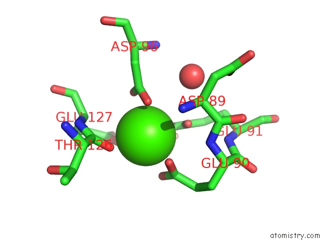
Mono view
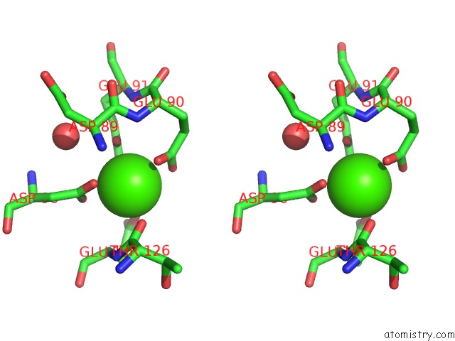
Stereo pair view
Mono view
Stereo pair view
A full contact list of Calcium with other atoms in the Ca binding
site number 1 of Crystal Structure of the N-Terminal Signalling Domain of Human Dhh with Calcium within 5.0Å range:
|
Calcium binding site 2 out of 2 in 2wfr
Go back to
Calcium binding site 2 out
of 2 in the Crystal Structure of the N-Terminal Signalling Domain of Human Dhh with Calcium
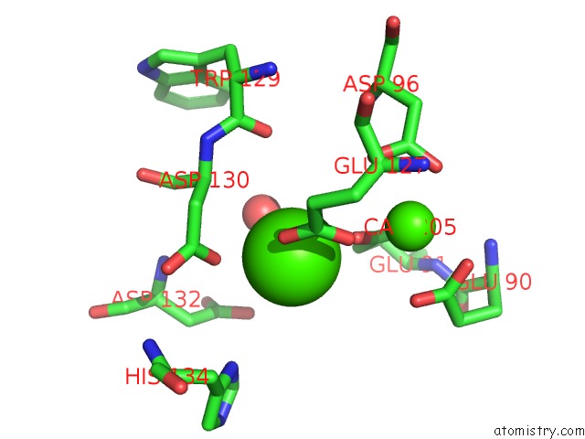
Mono view
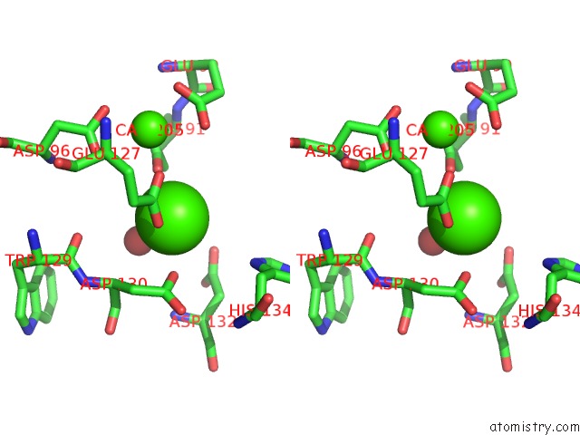
Stereo pair view
Mono view
Stereo pair view
A full contact list of Calcium with other atoms in the Ca binding
site number 2 of Crystal Structure of the N-Terminal Signalling Domain of Human Dhh with Calcium within 5.0Å range:
|
Reference:
B.Bishop,
A.R.Aricescu,
K.Harlos,
C.A.O'callaghan,
E.Y.Jones,
C.Siebold.
Structural Insights Into Hedgehog Ligand Sequestration By the Human Hedgehog-Interacting Protein Hip Nat.Struct.Mol.Biol. V. 16 698 2009.
ISSN: ISSN 1545-9993
PubMed: 19561611
DOI: 10.1038/NSMB.1607
Page generated: Tue Jul 8 09:04:53 2025
ISSN: ISSN 1545-9993
PubMed: 19561611
DOI: 10.1038/NSMB.1607
Last articles
Cl in 5LA5Cl in 5L9J
Cl in 5L9Z
Cl in 5L7I
Cl in 5L8D
Cl in 5L7U
Cl in 5L7P
Cl in 5L6I
Cl in 5L6H
Cl in 5L7F