Calcium »
PDB 2whv-2wtf »
2wnd »
Calcium in PDB 2wnd: Structure of An S100A7 Triple Mutant
Protein crystallography data
The structure of Structure of An S100A7 Triple Mutant, PDB code: 2wnd
was solved by
N.R.West,
B.Farnell,
P.H.Watson,
M.J.Boulanger,
with X-Ray Crystallography technique. A brief refinement statistics is given in the table below:
| Resolution Low / High (Å) | 24.37 / 1.60 |
| Space group | I 41 2 2 |
| Cell size a, b, c (Å), α, β, γ (°) | 68.930, 68.930, 92.520, 90.00, 90.00, 90.00 |
| R / Rfree (%) | 21.583 / 25.003 |
Other elements in 2wnd:
The structure of Structure of An S100A7 Triple Mutant also contains other interesting chemical elements:
| Zinc | (Zn) | 1 atom |
Calcium Binding Sites:
The binding sites of Calcium atom in the Structure of An S100A7 Triple Mutant
(pdb code 2wnd). This binding sites where shown within
5.0 Angstroms radius around Calcium atom.
In total only one binding site of Calcium was determined in the Structure of An S100A7 Triple Mutant, PDB code: 2wnd:
In total only one binding site of Calcium was determined in the Structure of An S100A7 Triple Mutant, PDB code: 2wnd:
Calcium binding site 1 out of 1 in 2wnd
Go back to
Calcium binding site 1 out
of 1 in the Structure of An S100A7 Triple Mutant
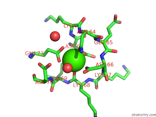
Mono view
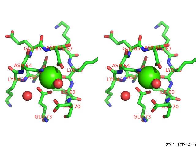
Stereo pair view
Mono view
Stereo pair view
A full contact list of Calcium with other atoms in the Ca binding
site number 1 of Structure of An S100A7 Triple Mutant within 5.0Å range:
|
Reference:
N.R.West,
B.Farnell,
J.I.Murray,
F.Hof,
P.H.Watson,
M.J.Boulanger.
Structural and Functional Characterization of A Triple Mutant Form of S100A7 Defective For JAB1 Binding. Protein Sci. V. 18 2615 2009.
ISSN: ISSN 0961-8368
PubMed: 19844956
DOI: 10.1002/PRO.274
Page generated: Fri Jul 12 18:45:15 2024
ISSN: ISSN 0961-8368
PubMed: 19844956
DOI: 10.1002/PRO.274
Last articles
Zn in 9J0NZn in 9J0O
Zn in 9J0P
Zn in 9FJX
Zn in 9EKB
Zn in 9C0F
Zn in 9CAH
Zn in 9CH0
Zn in 9CH3
Zn in 9CH1