Calcium »
PDB 2whv-2wtf »
2wos »
Calcium in PDB 2wos: Structure of Human S100A7 in Complex with 2,6 Ans
Protein crystallography data
The structure of Structure of Human S100A7 in Complex with 2,6 Ans, PDB code: 2wos
was solved by
R.Leon,
J.I.Murray,
G.Cragg,
B.Farnell,
T.C.Pace,
C.Bohne,
M.J.Boulanger,
F.Hof,
with X-Ray Crystallography technique. A brief refinement statistics is given in the table below:
| Resolution Low / High (Å) | 47.21 / 1.70 |
| Space group | P 43 21 2 |
| Cell size a, b, c (Å), α, β, γ (°) | 51.610, 51.610, 116.910, 90.00, 90.00, 90.00 |
| R / Rfree (%) | 18.07 / 21.023 |
Other elements in 2wos:
The structure of Structure of Human S100A7 in Complex with 2,6 Ans also contains other interesting chemical elements:
| Zinc | (Zn) | 1 atom |
Calcium Binding Sites:
The binding sites of Calcium atom in the Structure of Human S100A7 in Complex with 2,6 Ans
(pdb code 2wos). This binding sites where shown within
5.0 Angstroms radius around Calcium atom.
In total only one binding site of Calcium was determined in the Structure of Human S100A7 in Complex with 2,6 Ans, PDB code: 2wos:
In total only one binding site of Calcium was determined in the Structure of Human S100A7 in Complex with 2,6 Ans, PDB code: 2wos:
Calcium binding site 1 out of 1 in 2wos
Go back to
Calcium binding site 1 out
of 1 in the Structure of Human S100A7 in Complex with 2,6 Ans
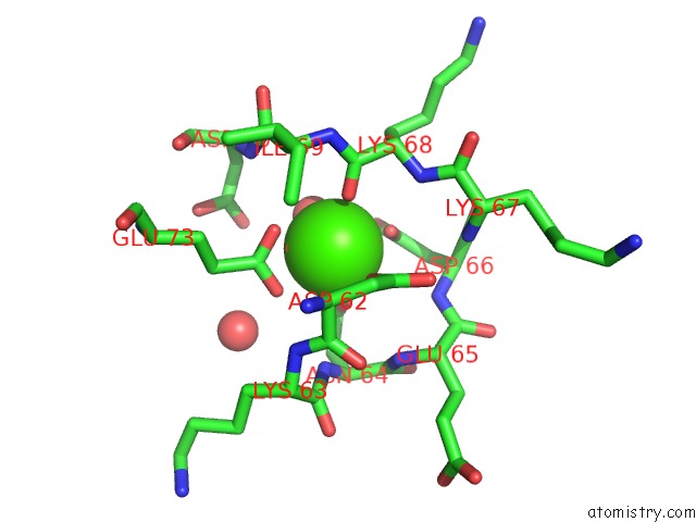
Mono view
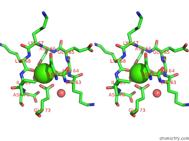
Stereo pair view
Mono view
Stereo pair view
A full contact list of Calcium with other atoms in the Ca binding
site number 1 of Structure of Human S100A7 in Complex with 2,6 Ans within 5.0Å range:
|
Reference:
R.Leon,
J.I.Murray,
G.Cragg,
B.Farnell,
N.R.West,
T.C.Pace,
P.H.Watson,
C.Bohne,
M.J.Boulanger,
F.Hof.
Identification and Characterization of Binding Sites on S100A7, A Participant in Cancer and Inflammation Pathways. Biochemistry V. 48 10591 2009.
ISSN: ISSN 0006-2960
PubMed: 19810752
DOI: 10.1021/BI901330G
Page generated: Tue Jul 8 09:10:39 2025
ISSN: ISSN 0006-2960
PubMed: 19810752
DOI: 10.1021/BI901330G
Last articles
Fe in 2YXOFe in 2YRS
Fe in 2YXC
Fe in 2YNM
Fe in 2YVJ
Fe in 2YP1
Fe in 2YU2
Fe in 2YU1
Fe in 2YQB
Fe in 2YOO