Calcium »
PDB 2xc5-2xon »
2xgq »
Calcium in PDB 2xgq: Structure of Yeast Dna Polymerase Eta in Complex with C8-N-Acetyl-2- Aminoanthracene Containing Dna
Enzymatic activity of Structure of Yeast Dna Polymerase Eta in Complex with C8-N-Acetyl-2- Aminoanthracene Containing Dna
All present enzymatic activity of Structure of Yeast Dna Polymerase Eta in Complex with C8-N-Acetyl-2- Aminoanthracene Containing Dna:
2.7.7.7;
2.7.7.7;
Protein crystallography data
The structure of Structure of Yeast Dna Polymerase Eta in Complex with C8-N-Acetyl-2- Aminoanthracene Containing Dna, PDB code: 2xgq
was solved by
S.Schneider,
S.Schorr,
T.Carell,
with X-Ray Crystallography technique. A brief refinement statistics is given in the table below:
| Resolution Low / High (Å) | 46.30 / 2.70 |
| Space group | P 41 21 2 |
| Cell size a, b, c (Å), α, β, γ (°) | 103.525, 103.525, 292.657, 90.00, 90.00, 90.00 |
| R / Rfree (%) | 21.7 / 26.4 |
Calcium Binding Sites:
The binding sites of Calcium atom in the Structure of Yeast Dna Polymerase Eta in Complex with C8-N-Acetyl-2- Aminoanthracene Containing Dna
(pdb code 2xgq). This binding sites where shown within
5.0 Angstroms radius around Calcium atom.
In total 7 binding sites of Calcium where determined in the Structure of Yeast Dna Polymerase Eta in Complex with C8-N-Acetyl-2- Aminoanthracene Containing Dna, PDB code: 2xgq:
Jump to Calcium binding site number: 1; 2; 3; 4; 5; 6; 7;
In total 7 binding sites of Calcium where determined in the Structure of Yeast Dna Polymerase Eta in Complex with C8-N-Acetyl-2- Aminoanthracene Containing Dna, PDB code: 2xgq:
Jump to Calcium binding site number: 1; 2; 3; 4; 5; 6; 7;
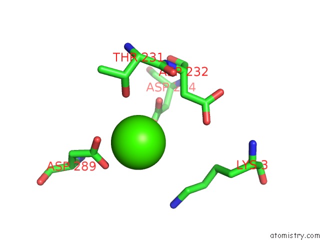
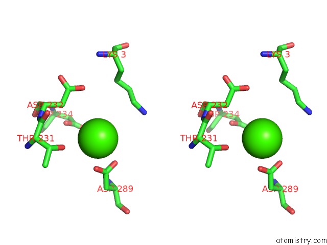
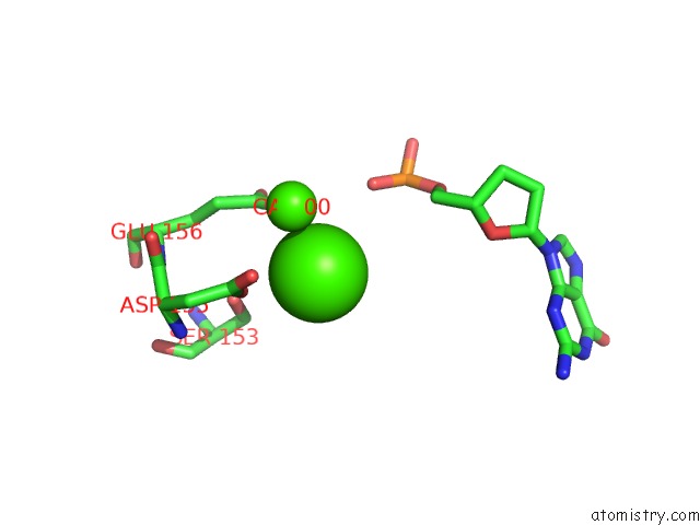
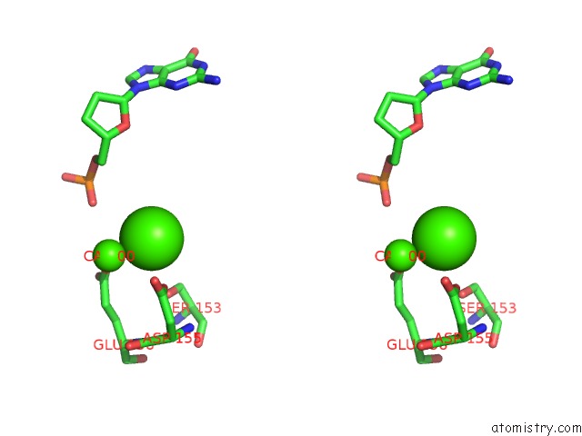
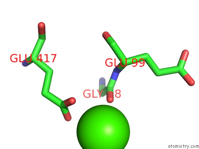
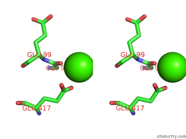
Calcium binding site 1 out of 7 in 2xgq
Go back to
Calcium binding site 1 out
of 7 in the Structure of Yeast Dna Polymerase Eta in Complex with C8-N-Acetyl-2- Aminoanthracene Containing Dna
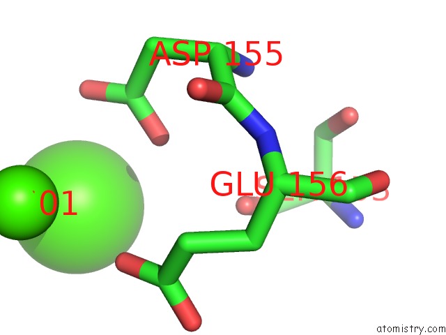
Mono view
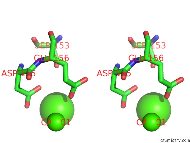
Stereo pair view
Mono view
Stereo pair view
A full contact list of Calcium with other atoms in the Ca binding
site number 1 of Structure of Yeast Dna Polymerase Eta in Complex with C8-N-Acetyl-2- Aminoanthracene Containing Dna within 5.0Å range:
|
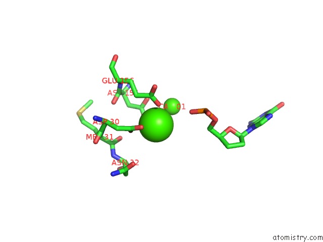
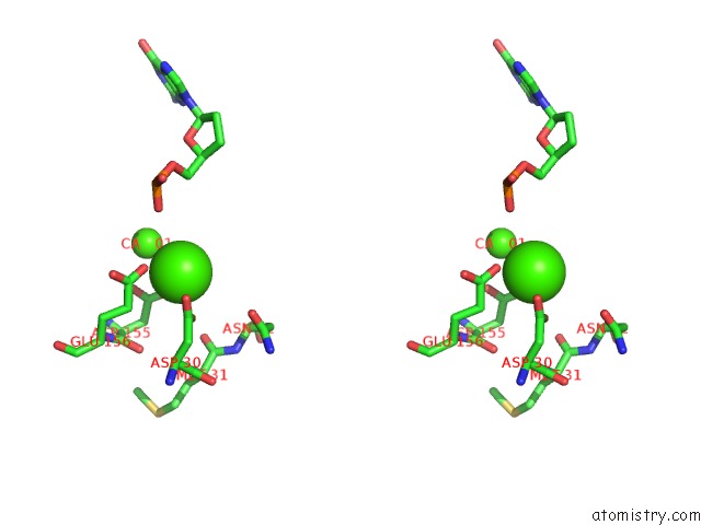
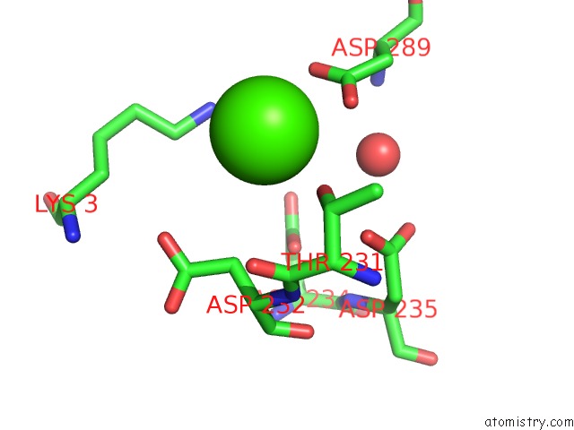
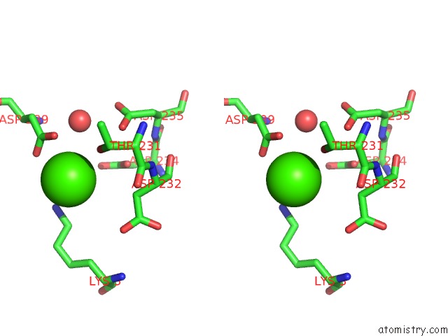
Calcium binding site 2 out of 7 in 2xgq
Go back to
Calcium binding site 2 out
of 7 in the Structure of Yeast Dna Polymerase Eta in Complex with C8-N-Acetyl-2- Aminoanthracene Containing Dna
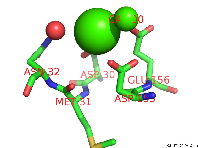
Mono view
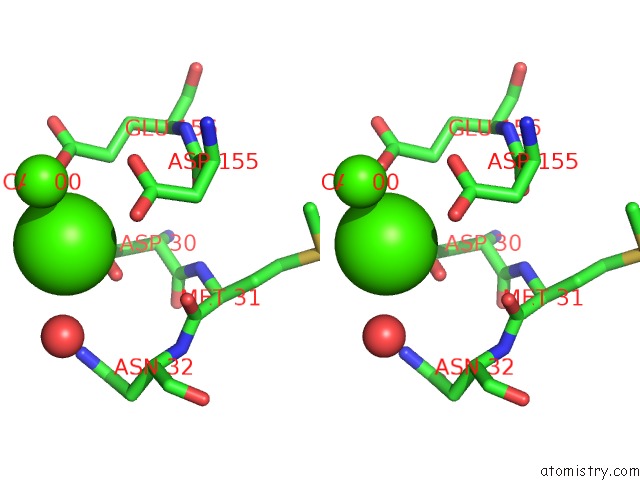
Stereo pair view
Mono view
Stereo pair view
A full contact list of Calcium with other atoms in the Ca binding
site number 2 of Structure of Yeast Dna Polymerase Eta in Complex with C8-N-Acetyl-2- Aminoanthracene Containing Dna within 5.0Å range:
|
Calcium binding site 3 out of 7 in 2xgq
Go back to
Calcium binding site 3 out
of 7 in the Structure of Yeast Dna Polymerase Eta in Complex with C8-N-Acetyl-2- Aminoanthracene Containing Dna
Mono view
Stereo pair view
Mono view
Stereo pair view
A full contact list of Calcium with other atoms in the Ca binding
site number 3 of Structure of Yeast Dna Polymerase Eta in Complex with C8-N-Acetyl-2- Aminoanthracene Containing Dna within 5.0Å range:
|
Calcium binding site 4 out of 7 in 2xgq
Go back to
Calcium binding site 4 out
of 7 in the Structure of Yeast Dna Polymerase Eta in Complex with C8-N-Acetyl-2- Aminoanthracene Containing Dna
Mono view
Stereo pair view
Mono view
Stereo pair view
A full contact list of Calcium with other atoms in the Ca binding
site number 4 of Structure of Yeast Dna Polymerase Eta in Complex with C8-N-Acetyl-2- Aminoanthracene Containing Dna within 5.0Å range:
|
Calcium binding site 5 out of 7 in 2xgq
Go back to
Calcium binding site 5 out
of 7 in the Structure of Yeast Dna Polymerase Eta in Complex with C8-N-Acetyl-2- Aminoanthracene Containing Dna
Mono view
Stereo pair view
Mono view
Stereo pair view
A full contact list of Calcium with other atoms in the Ca binding
site number 5 of Structure of Yeast Dna Polymerase Eta in Complex with C8-N-Acetyl-2- Aminoanthracene Containing Dna within 5.0Å range:
|
Calcium binding site 6 out of 7 in 2xgq
Go back to
Calcium binding site 6 out
of 7 in the Structure of Yeast Dna Polymerase Eta in Complex with C8-N-Acetyl-2- Aminoanthracene Containing Dna
Mono view
Stereo pair view
Mono view
Stereo pair view
A full contact list of Calcium with other atoms in the Ca binding
site number 6 of Structure of Yeast Dna Polymerase Eta in Complex with C8-N-Acetyl-2- Aminoanthracene Containing Dna within 5.0Å range:
|
Calcium binding site 7 out of 7 in 2xgq
Go back to
Calcium binding site 7 out
of 7 in the Structure of Yeast Dna Polymerase Eta in Complex with C8-N-Acetyl-2- Aminoanthracene Containing Dna
Mono view
Stereo pair view
Mono view
Stereo pair view
A full contact list of Calcium with other atoms in the Ca binding
site number 7 of Structure of Yeast Dna Polymerase Eta in Complex with C8-N-Acetyl-2- Aminoanthracene Containing Dna within 5.0Å range:
|
Reference:
S.Schorr,
S.Schneider,
K.Lammens,
K.P.Hopfner,
T.Carell.
Mechanism of Replication Blocking and Bypass of Y-Family Polymerase Eta By Bulky Acetylaminofluorene Dna Adducts. Proc.Natl.Acad.Sci.Usa V. 107 20720 2010.
ISSN: ISSN 0027-8424
PubMed: 21076032
DOI: 10.1073/PNAS.1008894107
Page generated: Tue Jul 8 09:22:30 2025
ISSN: ISSN 0027-8424
PubMed: 21076032
DOI: 10.1073/PNAS.1008894107
Last articles
Ca in 7G5UCa in 7G5V
Ca in 7G5W
Ca in 7G5T
Ca in 7G5R
Ca in 7G5S
Ca in 7G5Q
Ca in 7G5N
Ca in 7G5P
Ca in 7G5O