Calcium »
PDB 2xc5-2xon »
2xjm »
Calcium in PDB 2xjm: Crystal Structure of Streptococcus Suis Dpr with Cobalt
Protein crystallography data
The structure of Crystal Structure of Streptococcus Suis Dpr with Cobalt, PDB code: 2xjm
was solved by
T.Haikarainen,
A.Thanassoulas,
P.Stavros,
G.Nounesis,
S.Haataja,
A.C.Papageorgiou,
with X-Ray Crystallography technique. A brief refinement statistics is given in the table below:
| Resolution Low / High (Å) | 24.91 / 2.30 |
| Space group | P 21 21 21 |
| Cell size a, b, c (Å), α, β, γ (°) | 104.960, 137.420, 141.950, 90.00, 90.00, 90.00 |
| R / Rfree (%) | 17.7 / 24 |
Other elements in 2xjm:
The structure of Crystal Structure of Streptococcus Suis Dpr with Cobalt also contains other interesting chemical elements:
| Cobalt | (Co) | 12 atoms |
| Chlorine | (Cl) | 3 atoms |
Calcium Binding Sites:
The binding sites of Calcium atom in the Crystal Structure of Streptococcus Suis Dpr with Cobalt
(pdb code 2xjm). This binding sites where shown within
5.0 Angstroms radius around Calcium atom.
In total 8 binding sites of Calcium where determined in the Crystal Structure of Streptococcus Suis Dpr with Cobalt, PDB code: 2xjm:
Jump to Calcium binding site number: 1; 2; 3; 4; 5; 6; 7; 8;
In total 8 binding sites of Calcium where determined in the Crystal Structure of Streptococcus Suis Dpr with Cobalt, PDB code: 2xjm:
Jump to Calcium binding site number: 1; 2; 3; 4; 5; 6; 7; 8;
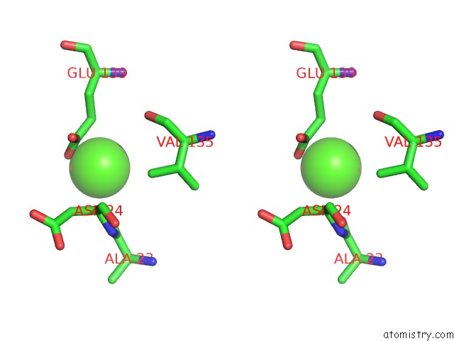
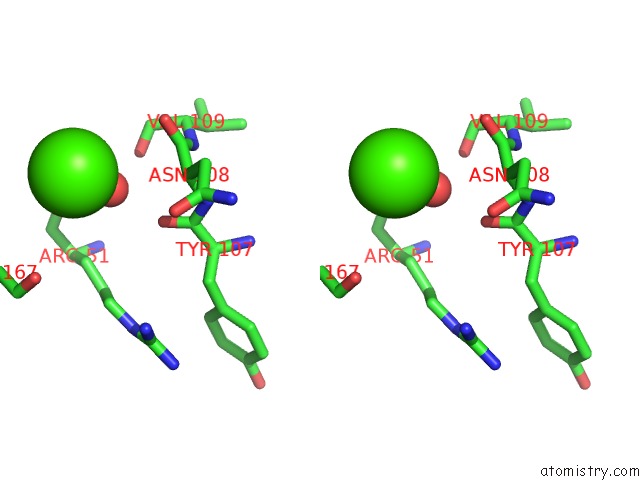
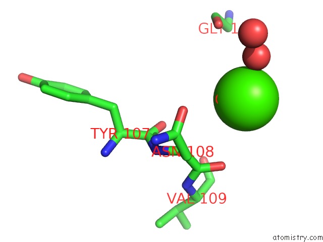
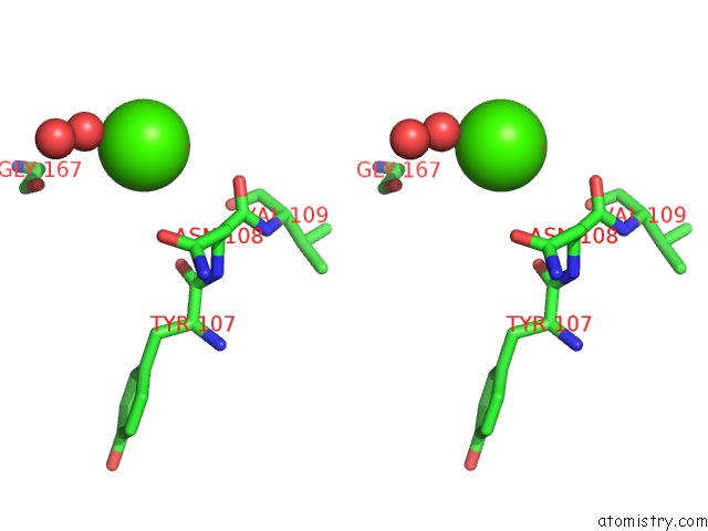
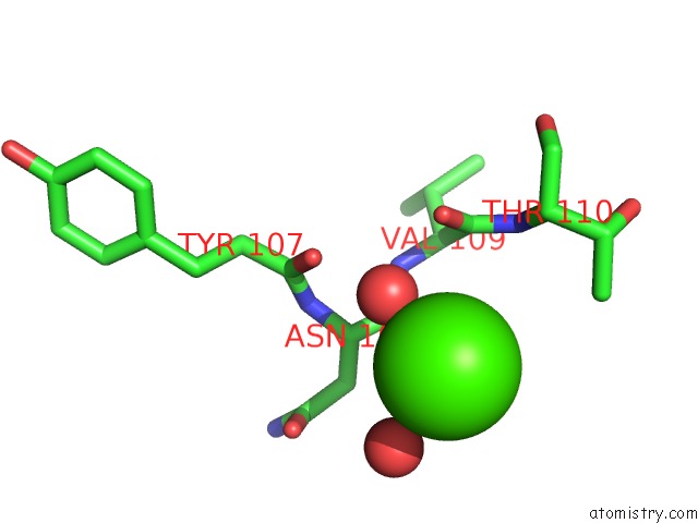
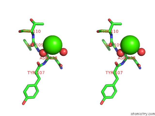
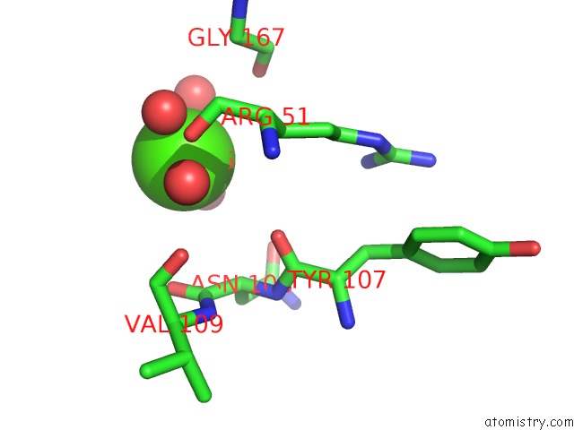
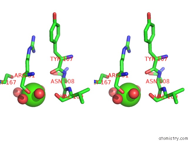
Calcium binding site 1 out of 8 in 2xjm
Go back to
Calcium binding site 1 out
of 8 in the Crystal Structure of Streptococcus Suis Dpr with Cobalt

Mono view
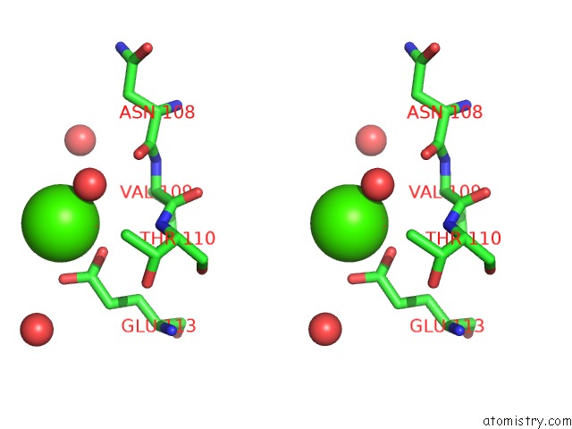
Stereo pair view
Mono view
Stereo pair view
A full contact list of Calcium with other atoms in the Ca binding
site number 1 of Crystal Structure of Streptococcus Suis Dpr with Cobalt within 5.0Å range:
|
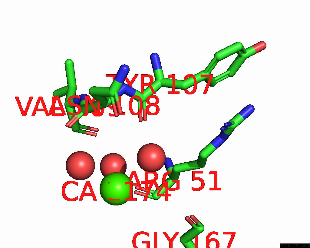
Calcium binding site 2 out of 8 in 2xjm
Go back to
Calcium binding site 2 out
of 8 in the Crystal Structure of Streptococcus Suis Dpr with Cobalt
Mono view
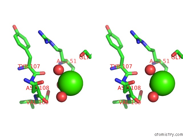
Stereo pair view
Mono view
Stereo pair view
A full contact list of Calcium with other atoms in the Ca binding
site number 2 of Crystal Structure of Streptococcus Suis Dpr with Cobalt within 5.0Å range:
|
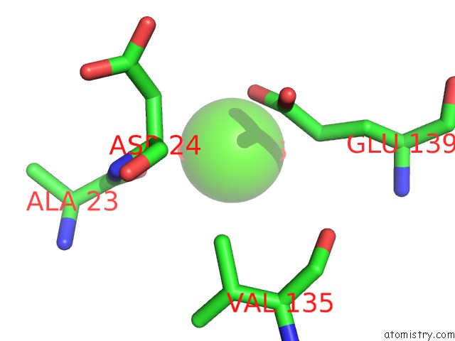
Calcium binding site 3 out of 8 in 2xjm
Go back to
Calcium binding site 3 out
of 8 in the Crystal Structure of Streptococcus Suis Dpr with Cobalt
Mono view
Stereo pair view
Mono view
Stereo pair view
A full contact list of Calcium with other atoms in the Ca binding
site number 3 of Crystal Structure of Streptococcus Suis Dpr with Cobalt within 5.0Å range:
|
Calcium binding site 4 out of 8 in 2xjm
Go back to
Calcium binding site 4 out
of 8 in the Crystal Structure of Streptococcus Suis Dpr with Cobalt
Mono view
Stereo pair view
Mono view
Stereo pair view
A full contact list of Calcium with other atoms in the Ca binding
site number 4 of Crystal Structure of Streptococcus Suis Dpr with Cobalt within 5.0Å range:
|
Calcium binding site 5 out of 8 in 2xjm
Go back to
Calcium binding site 5 out
of 8 in the Crystal Structure of Streptococcus Suis Dpr with Cobalt
Mono view
Stereo pair view
Mono view
Stereo pair view
A full contact list of Calcium with other atoms in the Ca binding
site number 5 of Crystal Structure of Streptococcus Suis Dpr with Cobalt within 5.0Å range:
|
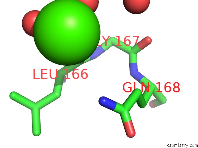
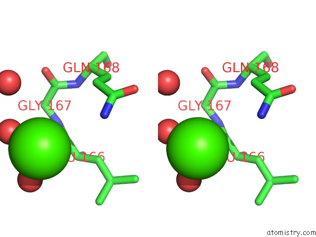
Calcium binding site 6 out of 8 in 2xjm
Go back to
Calcium binding site 6 out
of 8 in the Crystal Structure of Streptococcus Suis Dpr with Cobalt
Mono view
Stereo pair view
Mono view
Stereo pair view
A full contact list of Calcium with other atoms in the Ca binding
site number 6 of Crystal Structure of Streptococcus Suis Dpr with Cobalt within 5.0Å range:
|
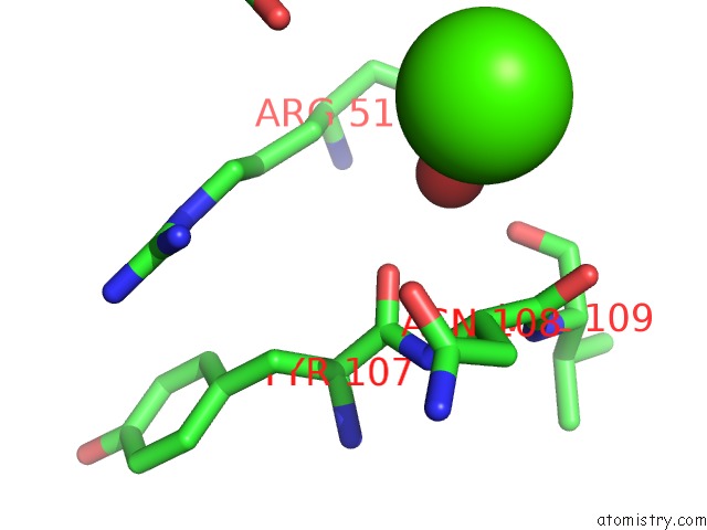
Calcium binding site 7 out of 8 in 2xjm
Go back to
Calcium binding site 7 out
of 8 in the Crystal Structure of Streptococcus Suis Dpr with Cobalt
Mono view
Stereo pair view
Mono view
Stereo pair view
A full contact list of Calcium with other atoms in the Ca binding
site number 7 of Crystal Structure of Streptococcus Suis Dpr with Cobalt within 5.0Å range:
|
Calcium binding site 8 out of 8 in 2xjm
Go back to
Calcium binding site 8 out
of 8 in the Crystal Structure of Streptococcus Suis Dpr with Cobalt
Mono view
Stereo pair view
Mono view
Stereo pair view
A full contact list of Calcium with other atoms in the Ca binding
site number 8 of Crystal Structure of Streptococcus Suis Dpr with Cobalt within 5.0Å range:
|
Reference:
T.Haikarainen,
A.Thanassoulas,
P.Stavros,
G.Nounesis,
S.Haataja,
A.C.Papageorgiou.
Structural and Thermodynamic Characterization of Metal Ion Binding in Streptococcus Suis Dpr. J.Mol.Biol. V. 405 448 2011.
ISSN: ISSN 0022-2836
PubMed: 21056572
DOI: 10.1016/J.JMB.2010.10.058
Page generated: Tue Jul 8 09:23:37 2025
ISSN: ISSN 0022-2836
PubMed: 21056572
DOI: 10.1016/J.JMB.2010.10.058
Last articles
Ca in 7ONGCa in 7OR0
Ca in 7OP2
Ca in 7OO7
Ca in 7OO5
Ca in 7O85
Ca in 7OIH
Ca in 7ONA
Ca in 7OMG
Ca in 7OKW