Calcium »
PDB 2xqg-2y6h »
2xtj »
Calcium in PDB 2xtj: The Crystal Structure of PCSK9 in Complex with 1D05 Fab
Protein crystallography data
The structure of The Crystal Structure of PCSK9 in Complex with 1D05 Fab, PDB code: 2xtj
was solved by
S.Di Marco,
C.Volpari,
A.Carfi,
with X-Ray Crystallography technique. A brief refinement statistics is given in the table below:
| Resolution Low / High (Å) | 40.00 / 2.70 |
| Space group | P 21 21 21 |
| Cell size a, b, c (Å), α, β, γ (°) | 66.589, 67.834, 250.860, 90.00, 90.00, 90.00 |
| R / Rfree (%) | 20 / 25.9 |
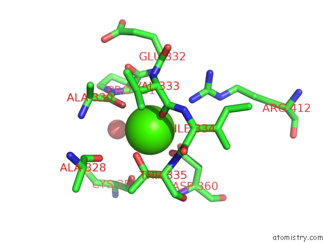
Calcium Binding Sites:
The binding sites of Calcium atom in the The Crystal Structure of PCSK9 in Complex with 1D05 Fab
(pdb code 2xtj). This binding sites where shown within
5.0 Angstroms radius around Calcium atom.
In total only one binding site of Calcium was determined in the The Crystal Structure of PCSK9 in Complex with 1D05 Fab, PDB code: 2xtj:
In total only one binding site of Calcium was determined in the The Crystal Structure of PCSK9 in Complex with 1D05 Fab, PDB code: 2xtj:
Calcium binding site 1 out of 1 in 2xtj
Go back to
Calcium binding site 1 out
of 1 in the The Crystal Structure of PCSK9 in Complex with 1D05 Fab
Mono view
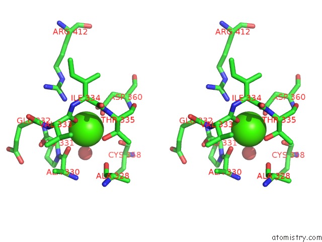
Stereo pair view
Mono view
Stereo pair view
A full contact list of Calcium with other atoms in the Ca binding
site number 1 of The Crystal Structure of PCSK9 in Complex with 1D05 Fab within 5.0Å range:
|
Reference:
Y.G.Ni,
S.Di Marco,
J.H.Condra,
L.B.Peterson,
W.Wang,
F.Wang,
S.Pandit,
H.A.Hammond,
R.Rosa,
R.T.Cummings,
D.D.Wood,
X.Liu,
M.J.Bottomley,
X.Shen,
R.M.Cubbon,
S.P.Wang,
D.G.Johns,
C.Volpari,
L.Hamuro,
J.Chin,
L.Huang,
J.Z.Zhao,
S.Vitelli,
P.Haytko,
D.Wisniewski,
L.J.Mitnaul,
C.P.Sparrow,
B.Hubbard,
A.Carfi,
A.Sitlani.
A PCSK9-Binding Antibody That Structurally Mimics the Egf(A) Domain of Ldl-Receptor Reduces Ldl Cholesterol in Vivo. J.Lipid Res. V. 52 78 2011.
ISSN: ISSN 0022-2275
PubMed: 20959675
DOI: 10.1194/JLR.M011445
Page generated: Tue Jul 8 09:29:00 2025
ISSN: ISSN 0022-2275
PubMed: 20959675
DOI: 10.1194/JLR.M011445
Last articles
Cl in 5HLOCl in 5HMA
Cl in 5HM3
Cl in 5HM0
Cl in 5HLY
Cl in 5HLV
Cl in 5HLW
Cl in 5HLN
Cl in 5HLL
Cl in 5HLS