Calcium »
PDB 2xqg-2y6h »
2xwg »
Calcium in PDB 2xwg: Crystal Structure of Sortase C-1 From Actinomyces Oris (Formerly Actinomyces Naeslundii)
Protein crystallography data
The structure of Crystal Structure of Sortase C-1 From Actinomyces Oris (Formerly Actinomyces Naeslundii), PDB code: 2xwg
was solved by
K.Persson,
with X-Ray Crystallography technique. A brief refinement statistics is given in the table below:
| Resolution Low / High (Å) | 43.67 / 2.40 |
| Space group | P 21 21 21 |
| Cell size a, b, c (Å), α, β, γ (°) | 104.070, 108.230, 143.200, 90.00, 90.00, 90.00 |
| R / Rfree (%) | 20.5 / 24.4 |
Calcium Binding Sites:
The binding sites of Calcium atom in the Crystal Structure of Sortase C-1 From Actinomyces Oris (Formerly Actinomyces Naeslundii)
(pdb code 2xwg). This binding sites where shown within
5.0 Angstroms radius around Calcium atom.
In total 4 binding sites of Calcium where determined in the Crystal Structure of Sortase C-1 From Actinomyces Oris (Formerly Actinomyces Naeslundii), PDB code: 2xwg:
Jump to Calcium binding site number: 1; 2; 3; 4;
In total 4 binding sites of Calcium where determined in the Crystal Structure of Sortase C-1 From Actinomyces Oris (Formerly Actinomyces Naeslundii), PDB code: 2xwg:
Jump to Calcium binding site number: 1; 2; 3; 4;
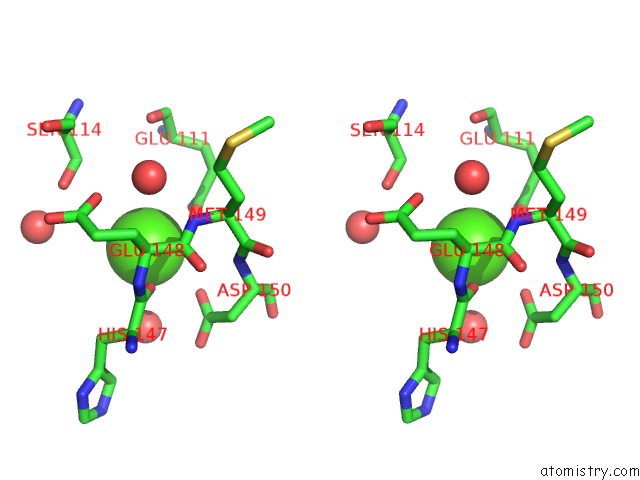
Calcium binding site 1 out of 4 in 2xwg
Go back to
Calcium binding site 1 out
of 4 in the Crystal Structure of Sortase C-1 From Actinomyces Oris (Formerly Actinomyces Naeslundii)

Mono view
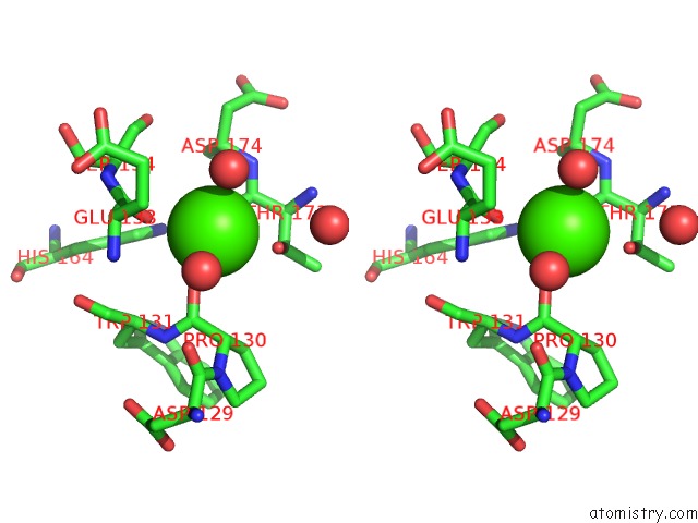
Stereo pair view
Mono view
Stereo pair view
A full contact list of Calcium with other atoms in the Ca binding
site number 1 of Crystal Structure of Sortase C-1 From Actinomyces Oris (Formerly Actinomyces Naeslundii) within 5.0Å range:
|
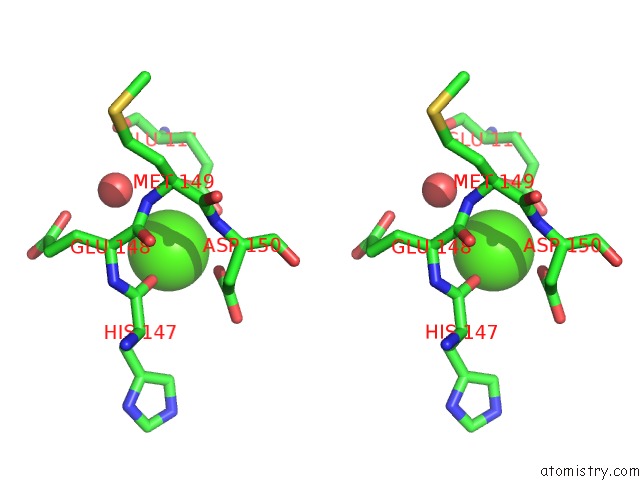
Calcium binding site 2 out of 4 in 2xwg
Go back to
Calcium binding site 2 out
of 4 in the Crystal Structure of Sortase C-1 From Actinomyces Oris (Formerly Actinomyces Naeslundii)
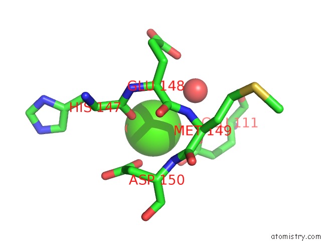
Mono view
Stereo pair view
Mono view
Stereo pair view
A full contact list of Calcium with other atoms in the Ca binding
site number 2 of Crystal Structure of Sortase C-1 From Actinomyces Oris (Formerly Actinomyces Naeslundii) within 5.0Å range:
|
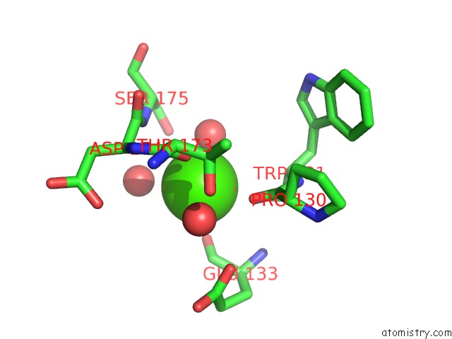
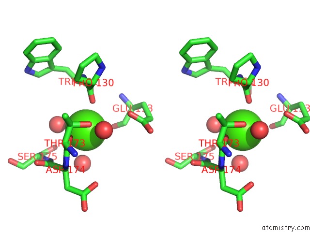
Calcium binding site 3 out of 4 in 2xwg
Go back to
Calcium binding site 3 out
of 4 in the Crystal Structure of Sortase C-1 From Actinomyces Oris (Formerly Actinomyces Naeslundii)
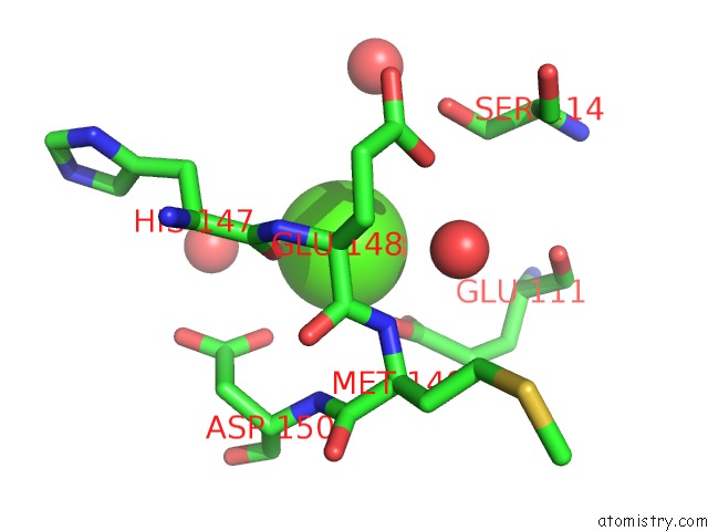
Mono view
Stereo pair view
Mono view
Stereo pair view
A full contact list of Calcium with other atoms in the Ca binding
site number 3 of Crystal Structure of Sortase C-1 From Actinomyces Oris (Formerly Actinomyces Naeslundii) within 5.0Å range:
|
Calcium binding site 4 out of 4 in 2xwg
Go back to
Calcium binding site 4 out
of 4 in the Crystal Structure of Sortase C-1 From Actinomyces Oris (Formerly Actinomyces Naeslundii)
Mono view
Stereo pair view
Mono view
Stereo pair view
A full contact list of Calcium with other atoms in the Ca binding
site number 4 of Crystal Structure of Sortase C-1 From Actinomyces Oris (Formerly Actinomyces Naeslundii) within 5.0Å range:
|
Reference:
K.Persson,
K.Persson.
N/A N/A.
ISSN: ISSN 0907-4449
PubMed: 21358052
DOI: 10.1107/S0907444911004215
Page generated: Fri Jul 12 19:16:28 2024
ISSN: ISSN 0907-4449
PubMed: 21358052
DOI: 10.1107/S0907444911004215
Last articles
Zn in 9MJ5Zn in 9HNW
Zn in 9G0L
Zn in 9FNE
Zn in 9DZN
Zn in 9E0I
Zn in 9D32
Zn in 9DAK
Zn in 8ZXC
Zn in 8ZUF