Calcium »
PDB 2xqg-2y6h »
2y6g »
Calcium in PDB 2y6g: Cellopentaose Binding Mutated (X-2 L110F) CBM4-2 Carbohydrate Binding Module From A Thermostable Rhodothermus Marinus Xylanase
Enzymatic activity of Cellopentaose Binding Mutated (X-2 L110F) CBM4-2 Carbohydrate Binding Module From A Thermostable Rhodothermus Marinus Xylanase
All present enzymatic activity of Cellopentaose Binding Mutated (X-2 L110F) CBM4-2 Carbohydrate Binding Module From A Thermostable Rhodothermus Marinus Xylanase:
3.2.1.8;
3.2.1.8;
Protein crystallography data
The structure of Cellopentaose Binding Mutated (X-2 L110F) CBM4-2 Carbohydrate Binding Module From A Thermostable Rhodothermus Marinus Xylanase, PDB code: 2y6g
was solved by
L.Von Schantz,
M.Hakansson,
D.T.Logan,
B.Walse,
J.Osterlin,
E.Nordberg-Karlsson,
M.Ohlin,
with X-Ray Crystallography technique. A brief refinement statistics is given in the table below:
| Resolution Low / High (Å) | 30.00 / 1.30 |
| Space group | P 21 21 21 |
| Cell size a, b, c (Å), α, β, γ (°) | 48.560, 49.720, 62.520, 90.00, 90.00, 90.00 |
| R / Rfree (%) | 13.2 / 18.3 |
Calcium Binding Sites:
The binding sites of Calcium atom in the Cellopentaose Binding Mutated (X-2 L110F) CBM4-2 Carbohydrate Binding Module From A Thermostable Rhodothermus Marinus Xylanase
(pdb code 2y6g). This binding sites where shown within
5.0 Angstroms radius around Calcium atom.
In total 2 binding sites of Calcium where determined in the Cellopentaose Binding Mutated (X-2 L110F) CBM4-2 Carbohydrate Binding Module From A Thermostable Rhodothermus Marinus Xylanase, PDB code: 2y6g:
Jump to Calcium binding site number: 1; 2;
In total 2 binding sites of Calcium where determined in the Cellopentaose Binding Mutated (X-2 L110F) CBM4-2 Carbohydrate Binding Module From A Thermostable Rhodothermus Marinus Xylanase, PDB code: 2y6g:
Jump to Calcium binding site number: 1; 2;
Calcium binding site 1 out of 2 in 2y6g
Go back to
Calcium binding site 1 out
of 2 in the Cellopentaose Binding Mutated (X-2 L110F) CBM4-2 Carbohydrate Binding Module From A Thermostable Rhodothermus Marinus Xylanase
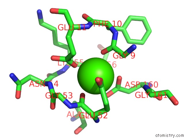
Mono view
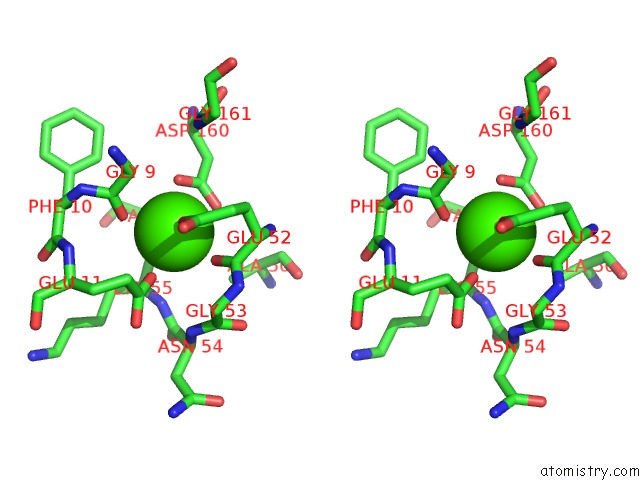
Stereo pair view
Mono view
Stereo pair view
A full contact list of Calcium with other atoms in the Ca binding
site number 1 of Cellopentaose Binding Mutated (X-2 L110F) CBM4-2 Carbohydrate Binding Module From A Thermostable Rhodothermus Marinus Xylanase within 5.0Å range:
|
Calcium binding site 2 out of 2 in 2y6g
Go back to
Calcium binding site 2 out
of 2 in the Cellopentaose Binding Mutated (X-2 L110F) CBM4-2 Carbohydrate Binding Module From A Thermostable Rhodothermus Marinus Xylanase
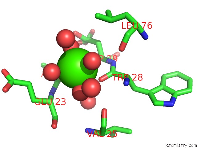
Mono view
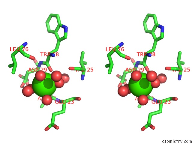
Stereo pair view
Mono view
Stereo pair view
A full contact list of Calcium with other atoms in the Ca binding
site number 2 of Cellopentaose Binding Mutated (X-2 L110F) CBM4-2 Carbohydrate Binding Module From A Thermostable Rhodothermus Marinus Xylanase within 5.0Å range:
|
Reference:
L.Von Schantz,
M.Hakansson,
D.T.Logan,
B.Walse,
J.Osterlin,
E.Nordberg-Karlsson,
M.Ohlin.
Structural Basis For Carbohydrate-Binding Specificity--A Comparative Assessment of Two Engineered Carbohydrate-Binding Modules. Glycobiology V. 22 948 2012.
ISSN: ESSN 1460-2423
PubMed: 22434778
DOI: 10.1093/GLYCOB/CWS063
Page generated: Fri Jul 12 19:22:36 2024
ISSN: ESSN 1460-2423
PubMed: 22434778
DOI: 10.1093/GLYCOB/CWS063
Last articles
Zn in 9MJ5Zn in 9HNW
Zn in 9G0L
Zn in 9FNE
Zn in 9DZN
Zn in 9E0I
Zn in 9D32
Zn in 9DAK
Zn in 8ZXC
Zn in 8ZUF