Calcium »
PDB 2y6j-2yhy »
2y6l »
Calcium in PDB 2y6l: Xylopentaose Binding X-2 Engineered Mutated CBM4-2 Carbohydrate Binding Module From A Thermostable Rhodothermus Marinus Xylanase
Enzymatic activity of Xylopentaose Binding X-2 Engineered Mutated CBM4-2 Carbohydrate Binding Module From A Thermostable Rhodothermus Marinus Xylanase
All present enzymatic activity of Xylopentaose Binding X-2 Engineered Mutated CBM4-2 Carbohydrate Binding Module From A Thermostable Rhodothermus Marinus Xylanase:
3.2.1.8;
3.2.1.8;
Protein crystallography data
The structure of Xylopentaose Binding X-2 Engineered Mutated CBM4-2 Carbohydrate Binding Module From A Thermostable Rhodothermus Marinus Xylanase, PDB code: 2y6l
was solved by
L.Von Schantz,
M.Hakansson,
D.T.Logan,
B.Walse,
J.Osterlin,
E.Nordberg-Karlsson,
M.Ohlin,
with X-Ray Crystallography technique. A brief refinement statistics is given in the table below:
| Resolution Low / High (Å) | 30.00 / 1.28 |
| Space group | P 21 21 21 |
| Cell size a, b, c (Å), α, β, γ (°) | 48.900, 49.480, 62.490, 90.00, 90.00, 90.00 |
| R / Rfree (%) | 13.2 / 18.3 |
Calcium Binding Sites:
The binding sites of Calcium atom in the Xylopentaose Binding X-2 Engineered Mutated CBM4-2 Carbohydrate Binding Module From A Thermostable Rhodothermus Marinus Xylanase
(pdb code 2y6l). This binding sites where shown within
5.0 Angstroms radius around Calcium atom.
In total 2 binding sites of Calcium where determined in the Xylopentaose Binding X-2 Engineered Mutated CBM4-2 Carbohydrate Binding Module From A Thermostable Rhodothermus Marinus Xylanase, PDB code: 2y6l:
Jump to Calcium binding site number: 1; 2;
In total 2 binding sites of Calcium where determined in the Xylopentaose Binding X-2 Engineered Mutated CBM4-2 Carbohydrate Binding Module From A Thermostable Rhodothermus Marinus Xylanase, PDB code: 2y6l:
Jump to Calcium binding site number: 1; 2;
Calcium binding site 1 out of 2 in 2y6l
Go back to
Calcium binding site 1 out
of 2 in the Xylopentaose Binding X-2 Engineered Mutated CBM4-2 Carbohydrate Binding Module From A Thermostable Rhodothermus Marinus Xylanase
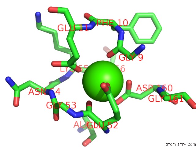
Mono view
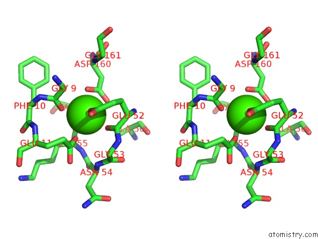
Stereo pair view
Mono view
Stereo pair view
A full contact list of Calcium with other atoms in the Ca binding
site number 1 of Xylopentaose Binding X-2 Engineered Mutated CBM4-2 Carbohydrate Binding Module From A Thermostable Rhodothermus Marinus Xylanase within 5.0Å range:
|
Calcium binding site 2 out of 2 in 2y6l
Go back to
Calcium binding site 2 out
of 2 in the Xylopentaose Binding X-2 Engineered Mutated CBM4-2 Carbohydrate Binding Module From A Thermostable Rhodothermus Marinus Xylanase
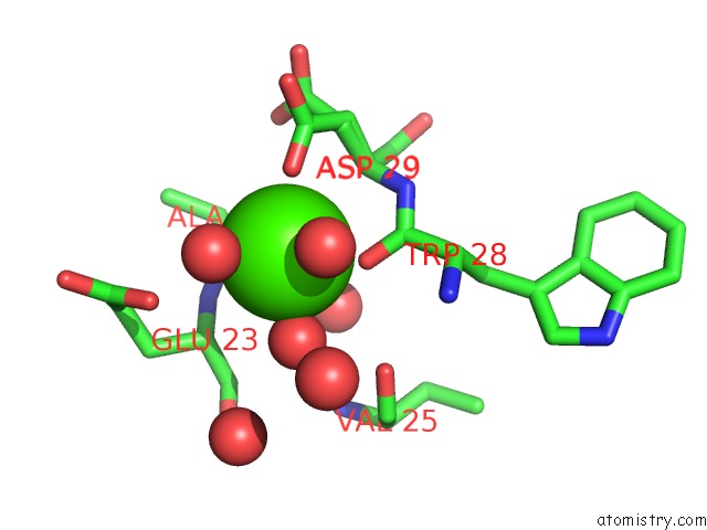
Mono view
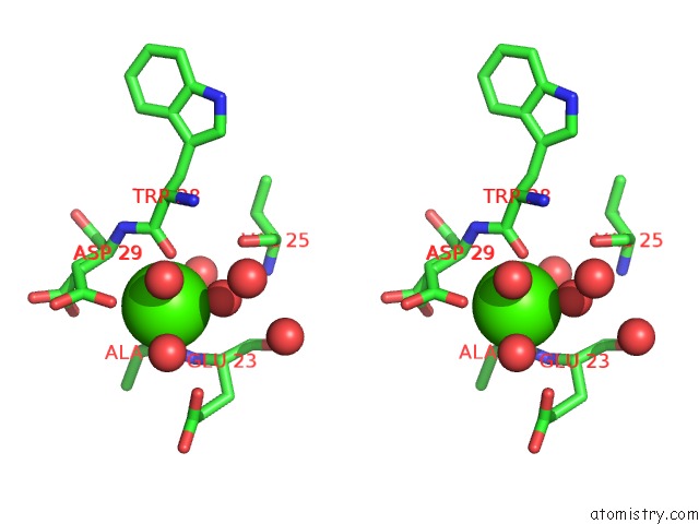
Stereo pair view
Mono view
Stereo pair view
A full contact list of Calcium with other atoms in the Ca binding
site number 2 of Xylopentaose Binding X-2 Engineered Mutated CBM4-2 Carbohydrate Binding Module From A Thermostable Rhodothermus Marinus Xylanase within 5.0Å range:
|
Reference:
L.Von Schantz,
M.Hakansson,
D.T.Logan,
B.Walse,
J.Osterlin,
E.Nordberg-Karlsson,
M.Ohlin.
Structural Basis For Carbohydrate-Binding Specificity--A Comparative Assessment of Two Engineered Carbohydrate-Binding Modules. Glycobiology V. 22 948 2012.
ISSN: ESSN 1460-2423
PubMed: 22434778
DOI: 10.1093/GLYCOB/CWS063
Page generated: Tue Jul 8 09:35:11 2025
ISSN: ESSN 1460-2423
PubMed: 22434778
DOI: 10.1093/GLYCOB/CWS063
Last articles
Cl in 8BX4Cl in 8BXS
Cl in 8BW1
Cl in 8BWX
Cl in 8BW7
Cl in 8BVP
Cl in 8BVS
Cl in 8BUQ
Cl in 8BU5
Cl in 8BU7