Calcium »
PDB 2y6j-2yhy »
2ydx »
Calcium in PDB 2ydx: Crystal Structure of Human S-Adenosylmethionine Synthetase 2, Beta Subunit
Protein crystallography data
The structure of Crystal Structure of Human S-Adenosylmethionine Synthetase 2, Beta Subunit, PDB code: 2ydx
was solved by
J.R.C.Muniz,
N.Shafqat,
A.C.W.Pike,
W.W.Yue,
M.Vollmar,
V.Papagriogriou,
A.Roos,
O.Gileadi,
F.Von Delft,
K.L.Kavanagh,
C.H.Arrowsmith,
A.M.Edwards,
J.Weigelt,
C.Bountra,
U.Oppermann,
with X-Ray Crystallography technique. A brief refinement statistics is given in the table below:
| Resolution Low / High (Å) | 58.96 / 2.80 |
| Space group | P 42 21 2 |
| Cell size a, b, c (Å), α, β, γ (°) | 163.389, 163.389, 252.882, 90.00, 90.00, 90.00 |
| R / Rfree (%) | 16.8 / 18.8 |
Calcium Binding Sites:
The binding sites of Calcium atom in the Crystal Structure of Human S-Adenosylmethionine Synthetase 2, Beta Subunit
(pdb code 2ydx). This binding sites where shown within
5.0 Angstroms radius around Calcium atom.
In total only one binding site of Calcium was determined in the Crystal Structure of Human S-Adenosylmethionine Synthetase 2, Beta Subunit, PDB code: 2ydx:
In total only one binding site of Calcium was determined in the Crystal Structure of Human S-Adenosylmethionine Synthetase 2, Beta Subunit, PDB code: 2ydx:
Calcium binding site 1 out of 1 in 2ydx
Go back to
Calcium binding site 1 out
of 1 in the Crystal Structure of Human S-Adenosylmethionine Synthetase 2, Beta Subunit
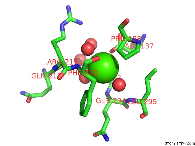
Mono view
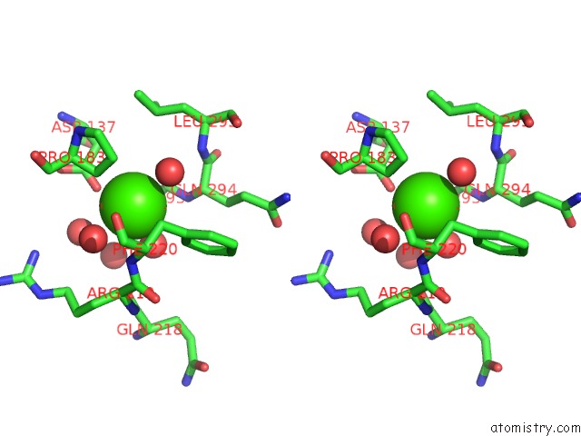
Stereo pair view
Mono view
Stereo pair view
A full contact list of Calcium with other atoms in the Ca binding
site number 1 of Crystal Structure of Human S-Adenosylmethionine Synthetase 2, Beta Subunit within 5.0Å range:
|
Reference:
N.Shafqat,
J.R.C.Muniz,
E.S.Pilka,
E.Papagrigoriou,
F.Von Delft,
U.Oppermann,
W.W.Yue.
Insight Into S-Adenosylmethionine Biosynthesis From the Crystal Structures of the Human Methionine Adenosyltransferase Catalytic and Regulatory Subunits. Biochem.J. V. 452 27 2013.
ISSN: ISSN 0264-6021
PubMed: 23425511
DOI: 10.1042/BJ20121580
Page generated: Fri Jul 12 19:28:25 2024
ISSN: ISSN 0264-6021
PubMed: 23425511
DOI: 10.1042/BJ20121580
Last articles
Zn in 9J0NZn in 9J0O
Zn in 9J0P
Zn in 9FJX
Zn in 9EKB
Zn in 9C0F
Zn in 9CAH
Zn in 9CH0
Zn in 9CH3
Zn in 9CH1