Calcium »
PDB 2y6j-2yhy »
2yft »
Calcium in PDB 2yft: Crystal Structure of Inulosucrase From Lactobacillus Johnsonii NCC533 in Complex with 1-Kestose
Enzymatic activity of Crystal Structure of Inulosucrase From Lactobacillus Johnsonii NCC533 in Complex with 1-Kestose
All present enzymatic activity of Crystal Structure of Inulosucrase From Lactobacillus Johnsonii NCC533 in Complex with 1-Kestose:
2.4.1.9;
2.4.1.9;
Protein crystallography data
The structure of Crystal Structure of Inulosucrase From Lactobacillus Johnsonii NCC533 in Complex with 1-Kestose, PDB code: 2yft
was solved by
T.Pijning,
M.A.Anwar,
H.Leemhuis,
S.Kralj,
L.Dijkhuizen,
B.W.Dijkstra,
with X-Ray Crystallography technique. A brief refinement statistics is given in the table below:
| Resolution Low / High (Å) | 43.89 / 1.85 |
| Space group | I 4 2 2 |
| Cell size a, b, c (Å), α, β, γ (°) | 171.204, 171.204, 115.003, 90.00, 90.00, 90.00 |
| R / Rfree (%) | 15.59 / 17.801 |
Calcium Binding Sites:
The binding sites of Calcium atom in the Crystal Structure of Inulosucrase From Lactobacillus Johnsonii NCC533 in Complex with 1-Kestose
(pdb code 2yft). This binding sites where shown within
5.0 Angstroms radius around Calcium atom.
In total only one binding site of Calcium was determined in the Crystal Structure of Inulosucrase From Lactobacillus Johnsonii NCC533 in Complex with 1-Kestose, PDB code: 2yft:
In total only one binding site of Calcium was determined in the Crystal Structure of Inulosucrase From Lactobacillus Johnsonii NCC533 in Complex with 1-Kestose, PDB code: 2yft:
Calcium binding site 1 out of 1 in 2yft
Go back to
Calcium binding site 1 out
of 1 in the Crystal Structure of Inulosucrase From Lactobacillus Johnsonii NCC533 in Complex with 1-Kestose
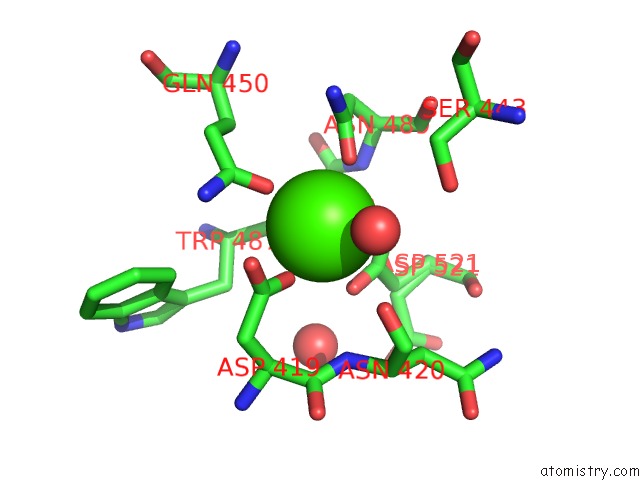
Mono view
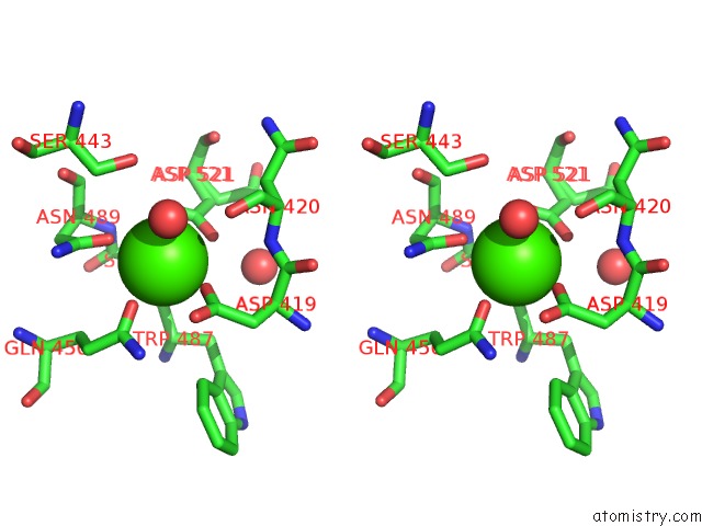
Stereo pair view
Mono view
Stereo pair view
A full contact list of Calcium with other atoms in the Ca binding
site number 1 of Crystal Structure of Inulosucrase From Lactobacillus Johnsonii NCC533 in Complex with 1-Kestose within 5.0Å range:
|
Reference:
T.Pijning,
M.A.Anwar,
M.Boger,
J.M.Dobruchowska,
H.Leemhuis,
S.Kralj,
L.Dijkhuizen,
B.W.Dijkstra.
Crystal Structure of Inulosucrase From Lactobacillus: Insights Into the Substrate Specificity and Product Specificity of GH68 Fructansucrases. J.Mol.Biol. V. 412 80 2011.
ISSN: ISSN 0022-2836
PubMed: 21801732
DOI: 10.1016/J.JMB.2011.07.031
Page generated: Fri Jul 12 19:29:27 2024
ISSN: ISSN 0022-2836
PubMed: 21801732
DOI: 10.1016/J.JMB.2011.07.031
Last articles
Zn in 9J0NZn in 9J0O
Zn in 9J0P
Zn in 9FJX
Zn in 9EKB
Zn in 9C0F
Zn in 9CAH
Zn in 9CH0
Zn in 9CH3
Zn in 9CH1