Calcium »
PDB 2z8x-2zp4 »
2zah »
Calcium in PDB 2zah: X-Ray Structure of Melon Necrotic Spot Virus
Protein crystallography data
The structure of X-Ray Structure of Melon Necrotic Spot Virus, PDB code: 2zah
was solved by
Y.Wada,
T.Tsukihara,
T.Omura,
with X-Ray Crystallography technique. A brief refinement statistics is given in the table below:
| Resolution Low / High (Å) | 187.50 / 2.81 |
| Space group | I 2 3 |
| Cell size a, b, c (Å), α, β, γ (°) | 375.004, 375.004, 375.004, 90.00, 90.00, 90.00 |
| R / Rfree (%) | 20.8 / 22.4 |
Calcium Binding Sites:
The binding sites of Calcium atom in the X-Ray Structure of Melon Necrotic Spot Virus
(pdb code 2zah). This binding sites where shown within
5.0 Angstroms radius around Calcium atom.
In total 4 binding sites of Calcium where determined in the X-Ray Structure of Melon Necrotic Spot Virus, PDB code: 2zah:
Jump to Calcium binding site number: 1; 2; 3; 4;
In total 4 binding sites of Calcium where determined in the X-Ray Structure of Melon Necrotic Spot Virus, PDB code: 2zah:
Jump to Calcium binding site number: 1; 2; 3; 4;
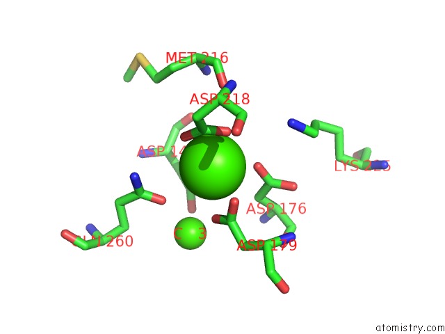
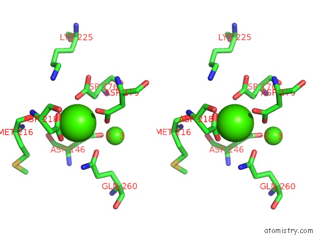
Calcium binding site 1 out of 4 in 2zah
Go back to
Calcium binding site 1 out
of 4 in the X-Ray Structure of Melon Necrotic Spot Virus
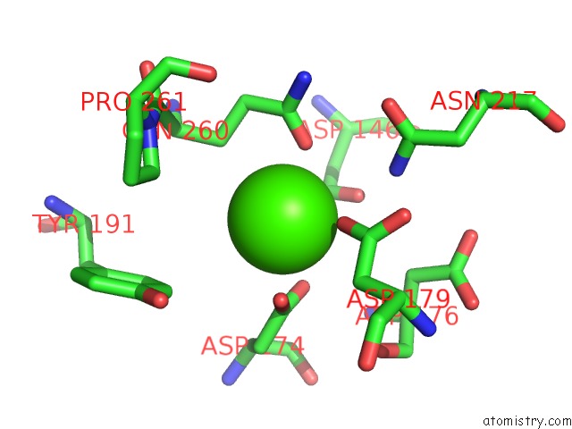
Mono view
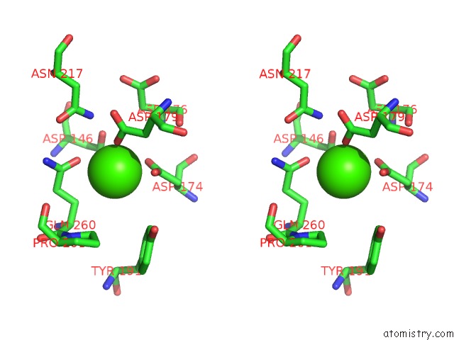
Stereo pair view
Mono view
Stereo pair view
A full contact list of Calcium with other atoms in the Ca binding
site number 1 of X-Ray Structure of Melon Necrotic Spot Virus within 5.0Å range:
|
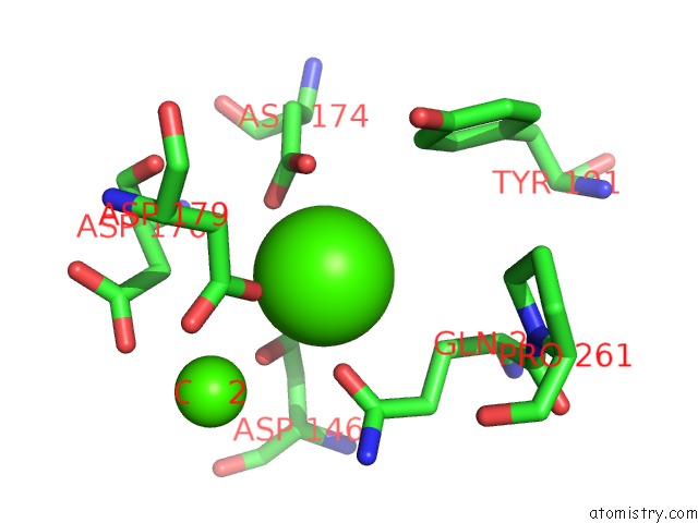
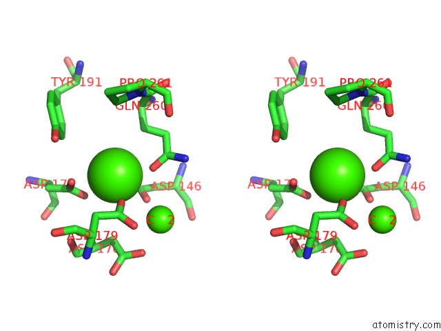
Calcium binding site 2 out of 4 in 2zah
Go back to
Calcium binding site 2 out
of 4 in the X-Ray Structure of Melon Necrotic Spot Virus
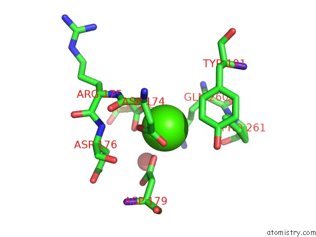
Mono view
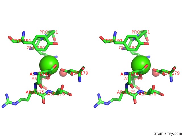
Stereo pair view
Mono view
Stereo pair view
A full contact list of Calcium with other atoms in the Ca binding
site number 2 of X-Ray Structure of Melon Necrotic Spot Virus within 5.0Å range:
|
Calcium binding site 3 out of 4 in 2zah
Go back to
Calcium binding site 3 out
of 4 in the X-Ray Structure of Melon Necrotic Spot Virus
Mono view
Stereo pair view
Mono view
Stereo pair view
A full contact list of Calcium with other atoms in the Ca binding
site number 3 of X-Ray Structure of Melon Necrotic Spot Virus within 5.0Å range:
|
Calcium binding site 4 out of 4 in 2zah
Go back to
Calcium binding site 4 out
of 4 in the X-Ray Structure of Melon Necrotic Spot Virus
Mono view
Stereo pair view
Mono view
Stereo pair view
A full contact list of Calcium with other atoms in the Ca binding
site number 4 of X-Ray Structure of Melon Necrotic Spot Virus within 5.0Å range:
|
Reference:
Y.Wada,
H.Tanaka,
E.Yamashita,
C.Kubo,
T.Ichiki-Uehara,
E.Nakazono-Nagaoka,
T.Omura,
T.Tsukihara.
X-Ray Structure of Melon Necrotic Spot Virus Acta Crystallogr.,Sect.F V. 64 8 2008.
ISSN: ESSN 1744-3091
PubMed: 18097092
DOI: 10.1107/S1744309107066481
Page generated: Fri Jul 12 19:57:12 2024
ISSN: ESSN 1744-3091
PubMed: 18097092
DOI: 10.1107/S1744309107066481
Last articles
Zn in 9MJ5Zn in 9HNW
Zn in 9G0L
Zn in 9FNE
Zn in 9DZN
Zn in 9E0I
Zn in 9D32
Zn in 9DAK
Zn in 8ZXC
Zn in 8ZUF