Calcium »
PDB 2z8x-2zp4 »
2zkm »
Calcium in PDB 2zkm: Crystal Structure of Phospholipase C Beta 2
Enzymatic activity of Crystal Structure of Phospholipase C Beta 2
All present enzymatic activity of Crystal Structure of Phospholipase C Beta 2:
3.1.4.11;
3.1.4.11;
Protein crystallography data
The structure of Crystal Structure of Phospholipase C Beta 2, PDB code: 2zkm
was solved by
S.N.Hicks,
M.R.Jezyk,
S.Gershberg,
J.P.Seifert,
T.K.Harden,
J.Sondek,
with X-Ray Crystallography technique. A brief refinement statistics is given in the table below:
| Resolution Low / High (Å) | 19.45 / 1.62 |
| Space group | P 21 21 21 |
| Cell size a, b, c (Å), α, β, γ (°) | 80.404, 86.379, 147.447, 90.00, 90.00, 90.00 |
| R / Rfree (%) | 19.6 / 21.3 |
Calcium Binding Sites:
The binding sites of Calcium atom in the Crystal Structure of Phospholipase C Beta 2
(pdb code 2zkm). This binding sites where shown within
5.0 Angstroms radius around Calcium atom.
In total only one binding site of Calcium was determined in the Crystal Structure of Phospholipase C Beta 2, PDB code: 2zkm:
In total only one binding site of Calcium was determined in the Crystal Structure of Phospholipase C Beta 2, PDB code: 2zkm:
Calcium binding site 1 out of 1 in 2zkm
Go back to
Calcium binding site 1 out
of 1 in the Crystal Structure of Phospholipase C Beta 2
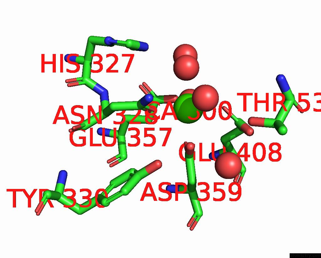
Mono view
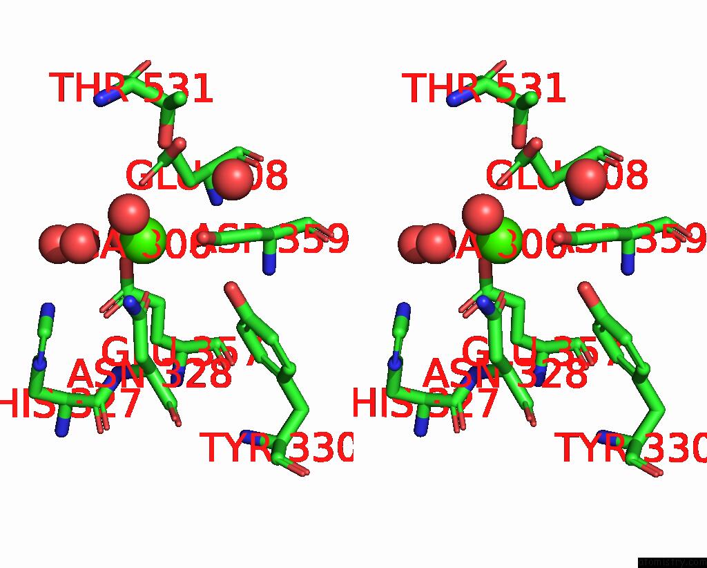
Stereo pair view
Mono view
Stereo pair view
A full contact list of Calcium with other atoms in the Ca binding
site number 1 of Crystal Structure of Phospholipase C Beta 2 within 5.0Å range:
|
Reference:
S.N.Hicks,
M.R.Jezyk,
S.Gershburg,
J.P.Seifert,
T.K.Harden,
J.Sondek.
General and Versatile Autoinhibition of Plc Isozymes Mol.Cell V. 31 383 2008.
ISSN: ISSN 1097-2765
PubMed: 18691970
DOI: 10.1016/J.MOLCEL.2008.06.018
Page generated: Fri Jul 12 20:03:59 2024
ISSN: ISSN 1097-2765
PubMed: 18691970
DOI: 10.1016/J.MOLCEL.2008.06.018
Last articles
Zn in 9J0NZn in 9J0O
Zn in 9J0P
Zn in 9FJX
Zn in 9EKB
Zn in 9C0F
Zn in 9CAH
Zn in 9CH0
Zn in 9CH3
Zn in 9CH1