Calcium »
PDB 2zp5-3a4h »
2zq0 »
Calcium in PDB 2zq0: Crystal Structure of Susb Complexed with Acarbose
Enzymatic activity of Crystal Structure of Susb Complexed with Acarbose
All present enzymatic activity of Crystal Structure of Susb Complexed with Acarbose:
3.2.1.20;
3.2.1.20;
Protein crystallography data
The structure of Crystal Structure of Susb Complexed with Acarbose, PDB code: 2zq0
was solved by
M.Yao,
I.Tanaka,
M.Kitamura,
with X-Ray Crystallography technique. A brief refinement statistics is given in the table below:
| Resolution Low / High (Å) | 20.00 / 1.60 |
| Space group | P 1 21 1 |
| Cell size a, b, c (Å), α, β, γ (°) | 75.717, 112.344, 102.467, 90.00, 100.61, 90.00 |
| R / Rfree (%) | 17.1 / 18.7 |
Calcium Binding Sites:
The binding sites of Calcium atom in the Crystal Structure of Susb Complexed with Acarbose
(pdb code 2zq0). This binding sites where shown within
5.0 Angstroms radius around Calcium atom.
In total 2 binding sites of Calcium where determined in the Crystal Structure of Susb Complexed with Acarbose, PDB code: 2zq0:
Jump to Calcium binding site number: 1; 2;
In total 2 binding sites of Calcium where determined in the Crystal Structure of Susb Complexed with Acarbose, PDB code: 2zq0:
Jump to Calcium binding site number: 1; 2;
Calcium binding site 1 out of 2 in 2zq0
Go back to
Calcium binding site 1 out
of 2 in the Crystal Structure of Susb Complexed with Acarbose
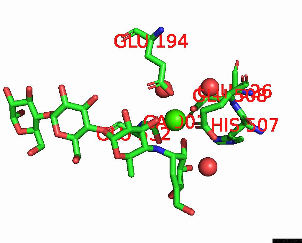
Mono view
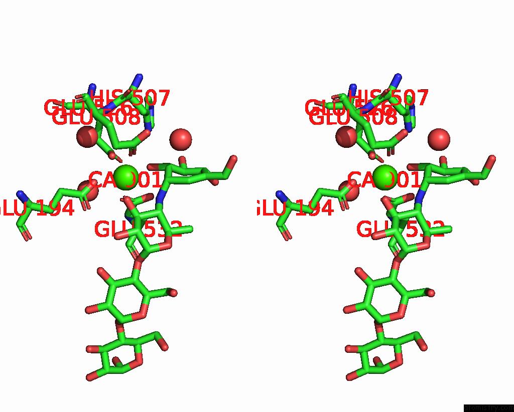
Stereo pair view
Mono view
Stereo pair view
A full contact list of Calcium with other atoms in the Ca binding
site number 1 of Crystal Structure of Susb Complexed with Acarbose within 5.0Å range:
|
Calcium binding site 2 out of 2 in 2zq0
Go back to
Calcium binding site 2 out
of 2 in the Crystal Structure of Susb Complexed with Acarbose
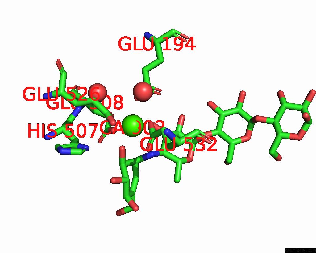
Mono view
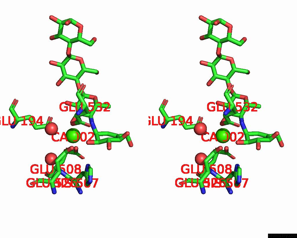
Stereo pair view
Mono view
Stereo pair view
A full contact list of Calcium with other atoms in the Ca binding
site number 2 of Crystal Structure of Susb Complexed with Acarbose within 5.0Å range:
|
Reference:
M.Kitamura,
M.Okuyama,
F.Tanzawa,
H.Mori,
Y.Kitago,
N.Watanabe,
A.Kimura,
I.Tanaka,
M.Yao.
Structural and Functional Analysis of A Glycoside Hydrolase Family 97 Enzyme From Bacteroides Thetaiotaomicron. J.Biol.Chem. V. 283 36328 2008.
ISSN: ISSN 0021-9258
PubMed: 18981178
DOI: 10.1074/JBC.M806115200
Page generated: Sat Jul 13 06:50:50 2024
ISSN: ISSN 0021-9258
PubMed: 18981178
DOI: 10.1074/JBC.M806115200
Last articles
Zn in 9J0NZn in 9J0O
Zn in 9J0P
Zn in 9FJX
Zn in 9EKB
Zn in 9C0F
Zn in 9CAH
Zn in 9CH0
Zn in 9CH3
Zn in 9CH1