Calcium »
PDB 2zp5-3a4h »
352d »
Calcium in PDB 352d: The Crystal Structure of A Parallel-Stranded Parallel- Stranded Guanine Tetraplex at 0.95 Angstrom Resolution
Protein crystallography data
The structure of The Crystal Structure of A Parallel-Stranded Parallel- Stranded Guanine Tetraplex at 0.95 Angstrom Resolution, PDB code: 352d
was solved by
K.Phillips,
Z.Dauter,
A.I.H.Murchie,
D.M.J.Lilley,
B.Luisi,
with X-Ray Crystallography technique. A brief refinement statistics is given in the table below:
| Resolution Low / High (Å) | N/A / 0.95 |
| Space group | P 1 |
| Cell size a, b, c (Å), α, β, γ (°) | 28.280, 34.780, 56.230, 74.31, 77.68, 89.81 |
| R / Rfree (%) | 15.2 / n/a |
Other elements in 352d:
The structure of The Crystal Structure of A Parallel-Stranded Parallel- Stranded Guanine Tetraplex at 0.95 Angstrom Resolution also contains other interesting chemical elements:
| Sodium | (Na) | 14 atoms |
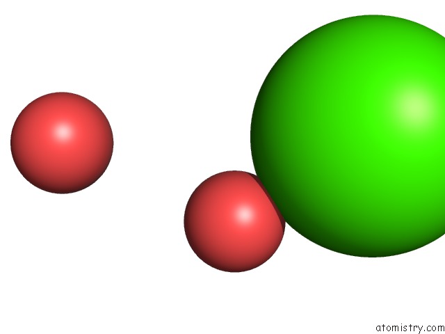
Calcium Binding Sites:
The binding sites of Calcium atom in the The Crystal Structure of A Parallel-Stranded Parallel- Stranded Guanine Tetraplex at 0.95 Angstrom Resolution
(pdb code 352d). This binding sites where shown within
5.0 Angstroms radius around Calcium atom.
In total 9 binding sites of Calcium where determined in the The Crystal Structure of A Parallel-Stranded Parallel- Stranded Guanine Tetraplex at 0.95 Angstrom Resolution, PDB code: 352d:
Jump to Calcium binding site number: 1; 2; 3; 4; 5; 6; 7; 8; 9;
In total 9 binding sites of Calcium where determined in the The Crystal Structure of A Parallel-Stranded Parallel- Stranded Guanine Tetraplex at 0.95 Angstrom Resolution, PDB code: 352d:
Jump to Calcium binding site number: 1; 2; 3; 4; 5; 6; 7; 8; 9;
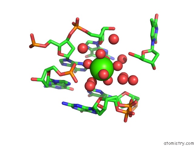
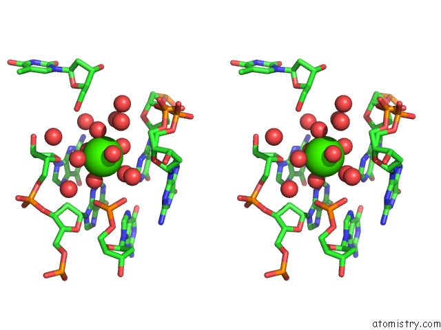
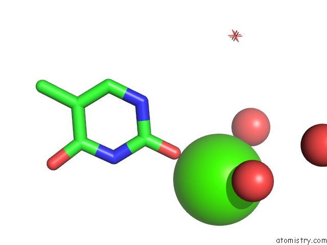
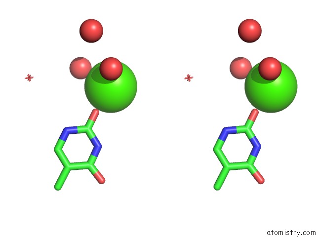
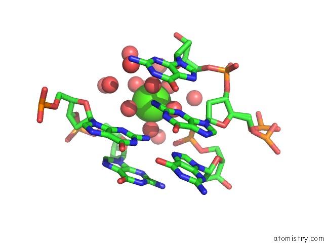
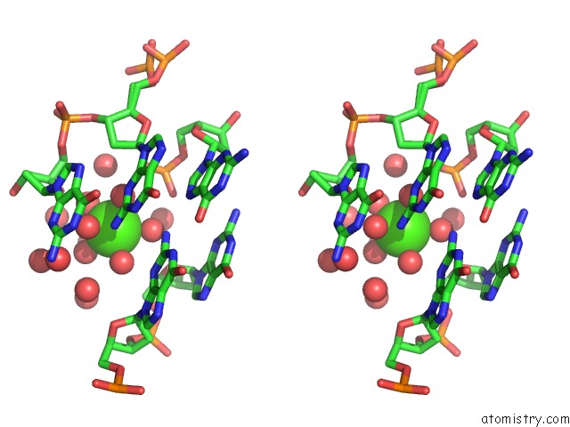
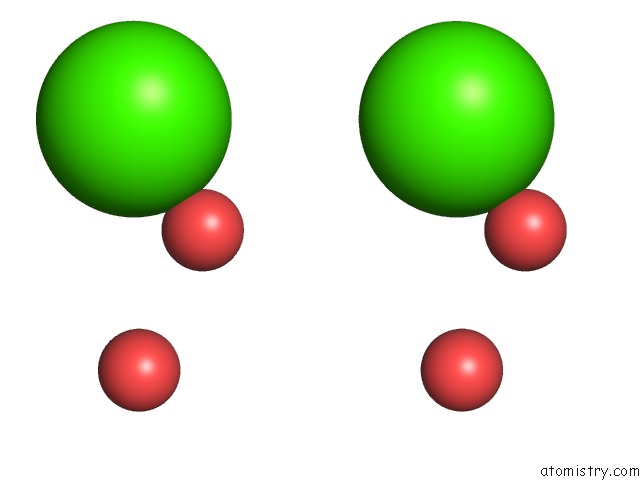
Calcium binding site 1 out of 9 in 352d
Go back to
Calcium binding site 1 out
of 9 in the The Crystal Structure of A Parallel-Stranded Parallel- Stranded Guanine Tetraplex at 0.95 Angstrom Resolution
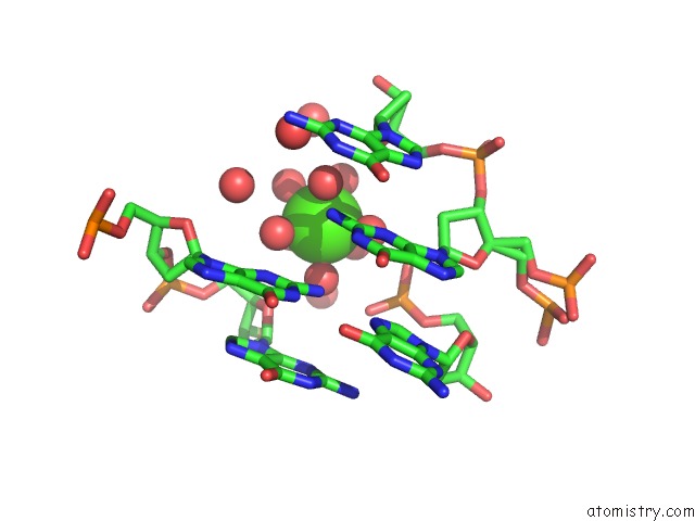
Mono view
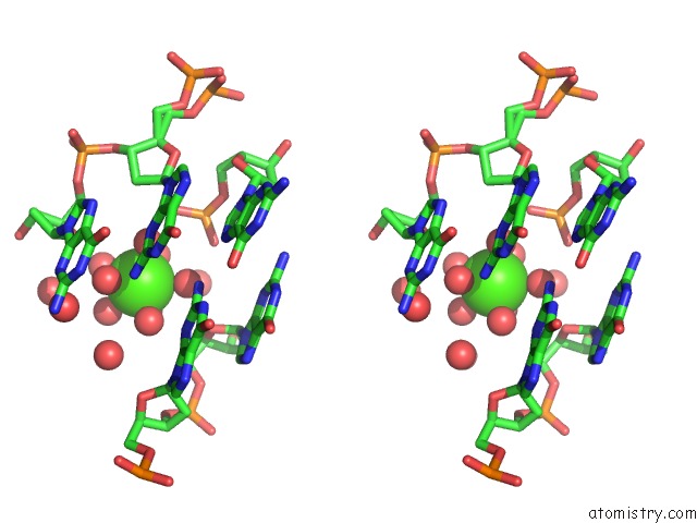
Stereo pair view
Mono view
Stereo pair view
A full contact list of Calcium with other atoms in the Ca binding
site number 1 of The Crystal Structure of A Parallel-Stranded Parallel- Stranded Guanine Tetraplex at 0.95 Angstrom Resolution within 5.0Å range:
|
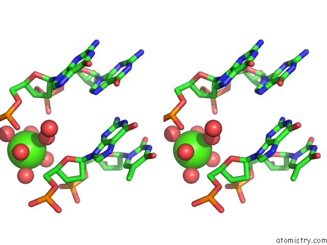
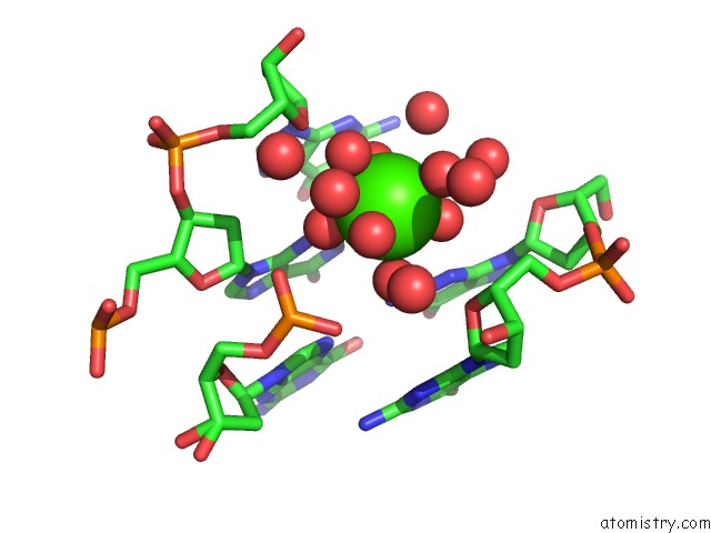
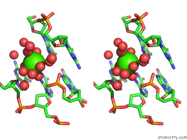
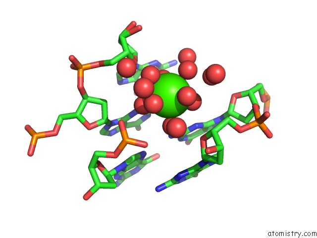
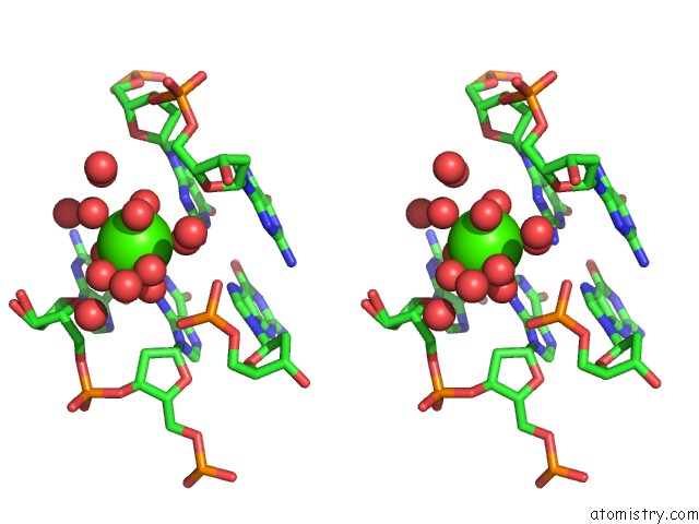
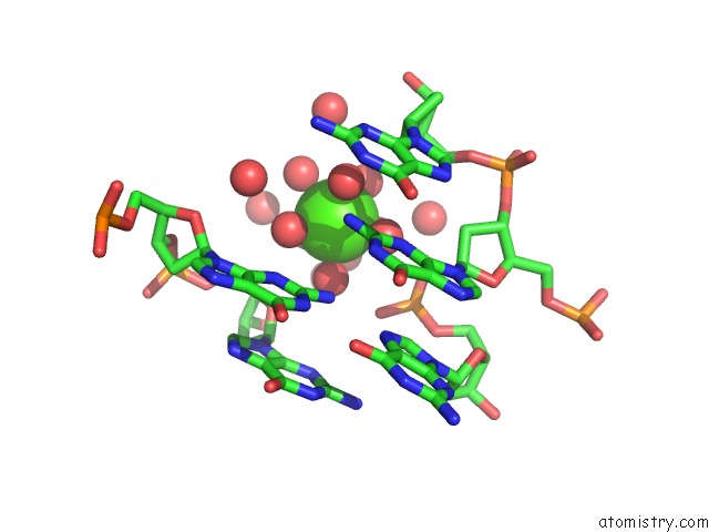
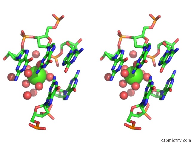
Calcium binding site 2 out of 9 in 352d
Go back to
Calcium binding site 2 out
of 9 in the The Crystal Structure of A Parallel-Stranded Parallel- Stranded Guanine Tetraplex at 0.95 Angstrom Resolution
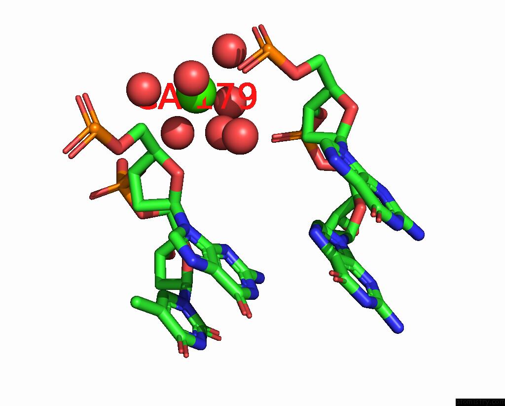
Mono view
Stereo pair view
Mono view
Stereo pair view
A full contact list of Calcium with other atoms in the Ca binding
site number 2 of The Crystal Structure of A Parallel-Stranded Parallel- Stranded Guanine Tetraplex at 0.95 Angstrom Resolution within 5.0Å range:
|
Calcium binding site 3 out of 9 in 352d
Go back to
Calcium binding site 3 out
of 9 in the The Crystal Structure of A Parallel-Stranded Parallel- Stranded Guanine Tetraplex at 0.95 Angstrom Resolution
Mono view
Stereo pair view
Mono view
Stereo pair view
A full contact list of Calcium with other atoms in the Ca binding
site number 3 of The Crystal Structure of A Parallel-Stranded Parallel- Stranded Guanine Tetraplex at 0.95 Angstrom Resolution within 5.0Å range:
|
Calcium binding site 4 out of 9 in 352d
Go back to
Calcium binding site 4 out
of 9 in the The Crystal Structure of A Parallel-Stranded Parallel- Stranded Guanine Tetraplex at 0.95 Angstrom Resolution
Mono view
Stereo pair view
Mono view
Stereo pair view
A full contact list of Calcium with other atoms in the Ca binding
site number 4 of The Crystal Structure of A Parallel-Stranded Parallel- Stranded Guanine Tetraplex at 0.95 Angstrom Resolution within 5.0Å range:
|
Calcium binding site 5 out of 9 in 352d
Go back to
Calcium binding site 5 out
of 9 in the The Crystal Structure of A Parallel-Stranded Parallel- Stranded Guanine Tetraplex at 0.95 Angstrom Resolution
Mono view
Stereo pair view
Mono view
Stereo pair view
A full contact list of Calcium with other atoms in the Ca binding
site number 5 of The Crystal Structure of A Parallel-Stranded Parallel- Stranded Guanine Tetraplex at 0.95 Angstrom Resolution within 5.0Å range:
|
Calcium binding site 6 out of 9 in 352d
Go back to
Calcium binding site 6 out
of 9 in the The Crystal Structure of A Parallel-Stranded Parallel- Stranded Guanine Tetraplex at 0.95 Angstrom Resolution
Mono view
Stereo pair view
Mono view
Stereo pair view
A full contact list of Calcium with other atoms in the Ca binding
site number 6 of The Crystal Structure of A Parallel-Stranded Parallel- Stranded Guanine Tetraplex at 0.95 Angstrom Resolution within 5.0Å range:
|
Calcium binding site 7 out of 9 in 352d
Go back to
Calcium binding site 7 out
of 9 in the The Crystal Structure of A Parallel-Stranded Parallel- Stranded Guanine Tetraplex at 0.95 Angstrom Resolution
Mono view
Stereo pair view
Mono view
Stereo pair view
A full contact list of Calcium with other atoms in the Ca binding
site number 7 of The Crystal Structure of A Parallel-Stranded Parallel- Stranded Guanine Tetraplex at 0.95 Angstrom Resolution within 5.0Å range:
|
Calcium binding site 8 out of 9 in 352d
Go back to
Calcium binding site 8 out
of 9 in the The Crystal Structure of A Parallel-Stranded Parallel- Stranded Guanine Tetraplex at 0.95 Angstrom Resolution
Mono view
Stereo pair view
Mono view
Stereo pair view
A full contact list of Calcium with other atoms in the Ca binding
site number 8 of The Crystal Structure of A Parallel-Stranded Parallel- Stranded Guanine Tetraplex at 0.95 Angstrom Resolution within 5.0Å range:
|
Calcium binding site 9 out of 9 in 352d
Go back to
Calcium binding site 9 out
of 9 in the The Crystal Structure of A Parallel-Stranded Parallel- Stranded Guanine Tetraplex at 0.95 Angstrom Resolution
Mono view
Stereo pair view
Mono view
Stereo pair view
A full contact list of Calcium with other atoms in the Ca binding
site number 9 of The Crystal Structure of A Parallel-Stranded Parallel- Stranded Guanine Tetraplex at 0.95 Angstrom Resolution within 5.0Å range:
|
Reference:
K.Phillips,
Z.Dauter,
A.I.Murchie,
D.M.Lilley,
B.Luisi.
The Crystal Structure of A Parallel-Stranded Guanine Tetraplex at 0.95 A Resolution. J.Mol.Biol. V. 273 171 1997.
ISSN: ISSN 0022-2836
PubMed: 9367755
DOI: 10.1006/JMBI.1997.1292
Page generated: Tue Jul 8 10:19:36 2025
ISSN: ISSN 0022-2836
PubMed: 9367755
DOI: 10.1006/JMBI.1997.1292
Last articles
Cl in 8BWKCl in 8BYB
Cl in 8BXT
Cl in 8BY9
Cl in 8BY0
Cl in 8BX4
Cl in 8BXS
Cl in 8BW1
Cl in 8BWX
Cl in 8BW7