Calcium »
PDB 2zp5-3a4h »
3a24 »
Calcium in PDB 3a24: Crystal Structure of BT1871 Retaining Glycosidase
Protein crystallography data
The structure of Crystal Structure of BT1871 Retaining Glycosidase, PDB code: 3a24
was solved by
M.Okuyama,
M.Kitamura,
H.Hondoh,
I.Tanaka,
M.Yao,
with X-Ray Crystallography technique. A brief refinement statistics is given in the table below:
| Resolution Low / High (Å) | 19.94 / 2.30 |
| Space group | I 2 2 2 |
| Cell size a, b, c (Å), α, β, γ (°) | 122.962, 167.100, 192.182, 90.00, 90.00, 90.00 |
| R / Rfree (%) | 18.6 / 21.8 |
Calcium Binding Sites:
The binding sites of Calcium atom in the Crystal Structure of BT1871 Retaining Glycosidase
(pdb code 3a24). This binding sites where shown within
5.0 Angstroms radius around Calcium atom.
In total 2 binding sites of Calcium where determined in the Crystal Structure of BT1871 Retaining Glycosidase, PDB code: 3a24:
Jump to Calcium binding site number: 1; 2;
In total 2 binding sites of Calcium where determined in the Crystal Structure of BT1871 Retaining Glycosidase, PDB code: 3a24:
Jump to Calcium binding site number: 1; 2;
Calcium binding site 1 out of 2 in 3a24
Go back to
Calcium binding site 1 out
of 2 in the Crystal Structure of BT1871 Retaining Glycosidase
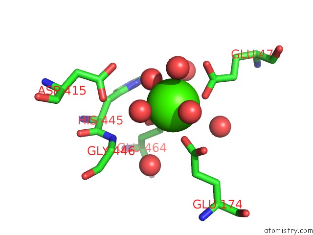
Mono view
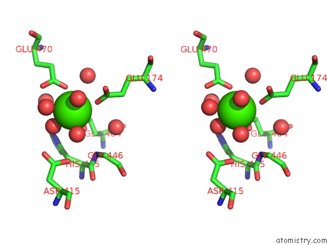
Stereo pair view
Mono view
Stereo pair view
A full contact list of Calcium with other atoms in the Ca binding
site number 1 of Crystal Structure of BT1871 Retaining Glycosidase within 5.0Å range:
|
Calcium binding site 2 out of 2 in 3a24
Go back to
Calcium binding site 2 out
of 2 in the Crystal Structure of BT1871 Retaining Glycosidase
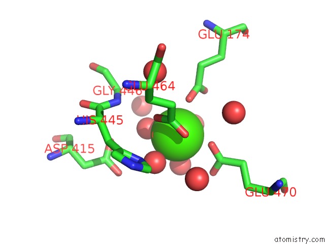
Mono view
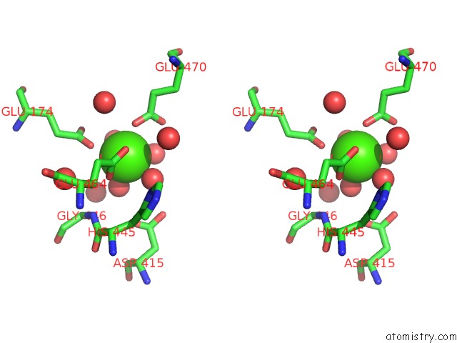
Stereo pair view
Mono view
Stereo pair view
A full contact list of Calcium with other atoms in the Ca binding
site number 2 of Crystal Structure of BT1871 Retaining Glycosidase within 5.0Å range:
|
Reference:
M.Okuyama,
M.Kitamura,
H.Hondoh,
M.S.Kang,
H.Mori,
A.Kimura,
I.Tanaka,
M.Yao.
Catalytic Mechanism of Retaining Alpha-Galactosidase Belonging to Glycoside Hydrolase Family 97. J.Mol.Biol. V. 392 1232 2009.
ISSN: ISSN 0022-2836
PubMed: 19646996
DOI: 10.1016/J.JMB.2009.07.068
Page generated: Sat Jul 13 07:06:29 2024
ISSN: ISSN 0022-2836
PubMed: 19646996
DOI: 10.1016/J.JMB.2009.07.068
Last articles
Zn in 9J0NZn in 9J0O
Zn in 9J0P
Zn in 9FJX
Zn in 9EKB
Zn in 9C0F
Zn in 9CAH
Zn in 9CH0
Zn in 9CH3
Zn in 9CH1