Calcium »
PDB 3a4u-3aas »
3a51 »
Calcium in PDB 3a51: Structure of Cytochrome P450 Vdh Mutant (Vdh-K1) Obtained By Directed Evolution with Bound 25-Hydroxyvitamin D3
Protein crystallography data
The structure of Structure of Cytochrome P450 Vdh Mutant (Vdh-K1) Obtained By Directed Evolution with Bound 25-Hydroxyvitamin D3, PDB code: 3a51
was solved by
Y.Yasutake,
Y.Fujii,
W.K.Cheon,
A.Arisawa,
T.Tamura,
with X-Ray Crystallography technique. A brief refinement statistics is given in the table below:
| Resolution Low / High (Å) | 45.60 / 2.00 |
| Space group | P 21 21 21 |
| Cell size a, b, c (Å), α, β, γ (°) | 77.173, 171.800, 189.143, 90.00, 90.00, 90.00 |
| R / Rfree (%) | 19.7 / 23.4 |
Other elements in 3a51:
The structure of Structure of Cytochrome P450 Vdh Mutant (Vdh-K1) Obtained By Directed Evolution with Bound 25-Hydroxyvitamin D3 also contains other interesting chemical elements:
| Iron | (Fe) | 5 atoms |
Calcium Binding Sites:
The binding sites of Calcium atom in the Structure of Cytochrome P450 Vdh Mutant (Vdh-K1) Obtained By Directed Evolution with Bound 25-Hydroxyvitamin D3
(pdb code 3a51). This binding sites where shown within
5.0 Angstroms radius around Calcium atom.
In total 6 binding sites of Calcium where determined in the Structure of Cytochrome P450 Vdh Mutant (Vdh-K1) Obtained By Directed Evolution with Bound 25-Hydroxyvitamin D3, PDB code: 3a51:
Jump to Calcium binding site number: 1; 2; 3; 4; 5; 6;
In total 6 binding sites of Calcium where determined in the Structure of Cytochrome P450 Vdh Mutant (Vdh-K1) Obtained By Directed Evolution with Bound 25-Hydroxyvitamin D3, PDB code: 3a51:
Jump to Calcium binding site number: 1; 2; 3; 4; 5; 6;
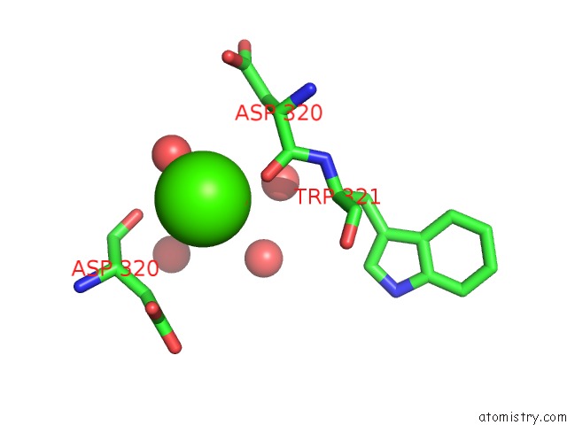
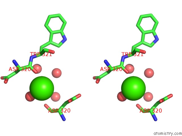
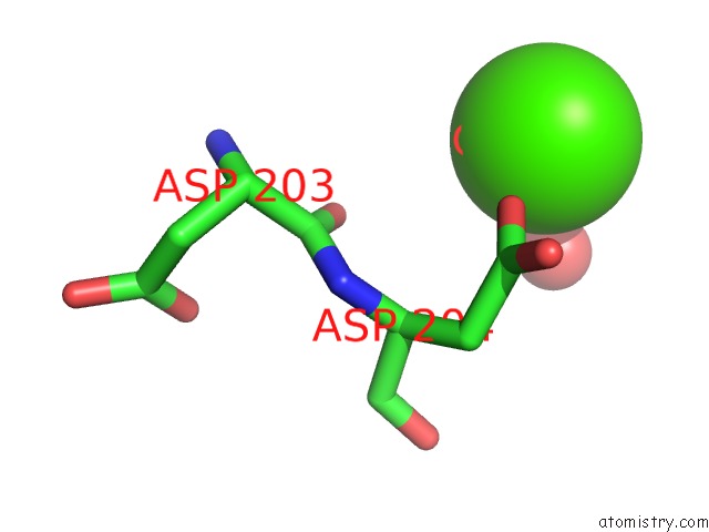
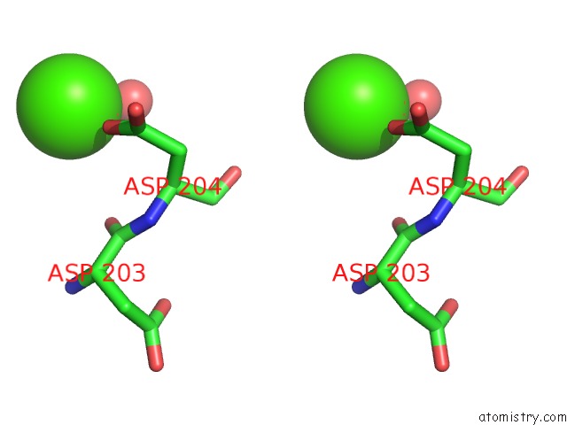
Calcium binding site 1 out of 6 in 3a51
Go back to
Calcium binding site 1 out
of 6 in the Structure of Cytochrome P450 Vdh Mutant (Vdh-K1) Obtained By Directed Evolution with Bound 25-Hydroxyvitamin D3
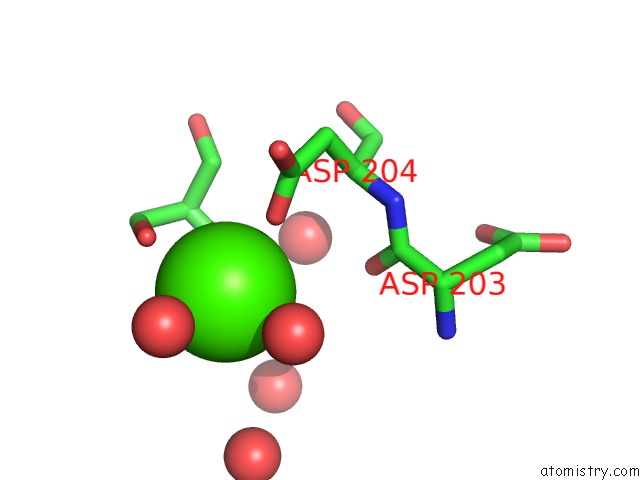
Mono view
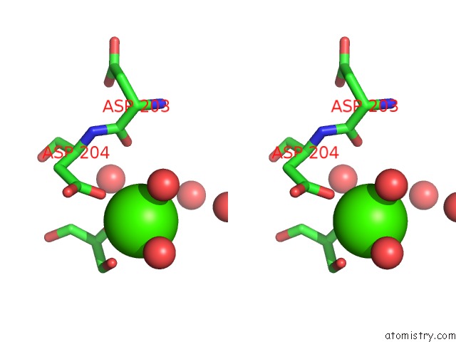
Stereo pair view
Mono view
Stereo pair view
A full contact list of Calcium with other atoms in the Ca binding
site number 1 of Structure of Cytochrome P450 Vdh Mutant (Vdh-K1) Obtained By Directed Evolution with Bound 25-Hydroxyvitamin D3 within 5.0Å range:
|
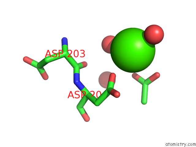
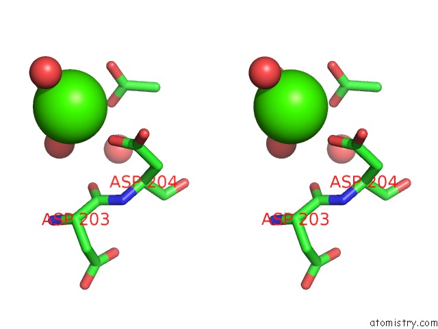
Calcium binding site 2 out of 6 in 3a51
Go back to
Calcium binding site 2 out
of 6 in the Structure of Cytochrome P450 Vdh Mutant (Vdh-K1) Obtained By Directed Evolution with Bound 25-Hydroxyvitamin D3
Mono view
Stereo pair view
Mono view
Stereo pair view
A full contact list of Calcium with other atoms in the Ca binding
site number 2 of Structure of Cytochrome P450 Vdh Mutant (Vdh-K1) Obtained By Directed Evolution with Bound 25-Hydroxyvitamin D3 within 5.0Å range:
|
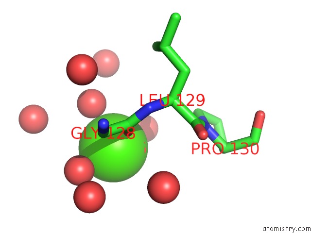
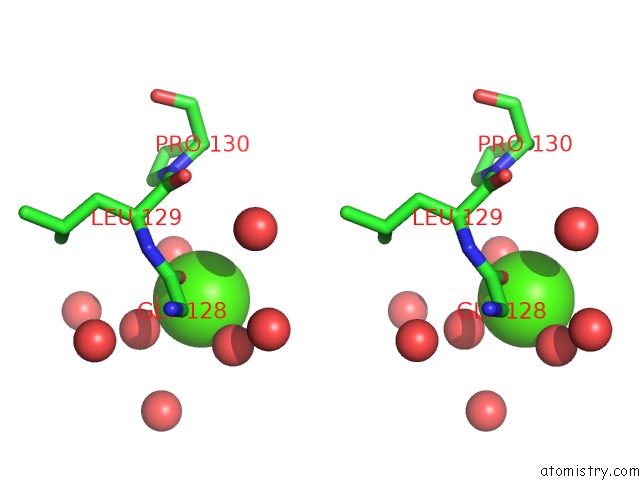
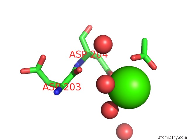
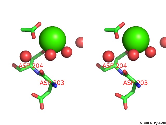
Calcium binding site 3 out of 6 in 3a51
Go back to
Calcium binding site 3 out
of 6 in the Structure of Cytochrome P450 Vdh Mutant (Vdh-K1) Obtained By Directed Evolution with Bound 25-Hydroxyvitamin D3
Mono view
Stereo pair view
Mono view
Stereo pair view
A full contact list of Calcium with other atoms in the Ca binding
site number 3 of Structure of Cytochrome P450 Vdh Mutant (Vdh-K1) Obtained By Directed Evolution with Bound 25-Hydroxyvitamin D3 within 5.0Å range:
|
Calcium binding site 4 out of 6 in 3a51
Go back to
Calcium binding site 4 out
of 6 in the Structure of Cytochrome P450 Vdh Mutant (Vdh-K1) Obtained By Directed Evolution with Bound 25-Hydroxyvitamin D3
Mono view
Stereo pair view
Mono view
Stereo pair view
A full contact list of Calcium with other atoms in the Ca binding
site number 4 of Structure of Cytochrome P450 Vdh Mutant (Vdh-K1) Obtained By Directed Evolution with Bound 25-Hydroxyvitamin D3 within 5.0Å range:
|
Calcium binding site 5 out of 6 in 3a51
Go back to
Calcium binding site 5 out
of 6 in the Structure of Cytochrome P450 Vdh Mutant (Vdh-K1) Obtained By Directed Evolution with Bound 25-Hydroxyvitamin D3
Mono view
Stereo pair view
Mono view
Stereo pair view
A full contact list of Calcium with other atoms in the Ca binding
site number 5 of Structure of Cytochrome P450 Vdh Mutant (Vdh-K1) Obtained By Directed Evolution with Bound 25-Hydroxyvitamin D3 within 5.0Å range:
|
Calcium binding site 6 out of 6 in 3a51
Go back to
Calcium binding site 6 out
of 6 in the Structure of Cytochrome P450 Vdh Mutant (Vdh-K1) Obtained By Directed Evolution with Bound 25-Hydroxyvitamin D3
Mono view
Stereo pair view
Mono view
Stereo pair view
A full contact list of Calcium with other atoms in the Ca binding
site number 6 of Structure of Cytochrome P450 Vdh Mutant (Vdh-K1) Obtained By Directed Evolution with Bound 25-Hydroxyvitamin D3 within 5.0Å range:
|
Reference:
Y.Yasutake,
Y.Fujii,
T.Nishioka,
W.K.Cheon,
A.Arisawa,
T.Tamura.
Structural Evidence For Enhancement of Sequential Vitamin D3 Hydroxylation Activities By Directed Evolution of Cytochrome P450 Vitamin D3 Hydroxylase J.Biol.Chem. V. 285 31193 2010.
ISSN: ISSN 0021-9258
PubMed: 20667833
DOI: 10.1074/JBC.M110.147009
Page generated: Sat Jul 13 07:11:30 2024
ISSN: ISSN 0021-9258
PubMed: 20667833
DOI: 10.1074/JBC.M110.147009
Last articles
Zn in 9J0NZn in 9J0O
Zn in 9J0P
Zn in 9FJX
Zn in 9EKB
Zn in 9C0F
Zn in 9CAH
Zn in 9CH0
Zn in 9CH3
Zn in 9CH1