Calcium »
PDB 3a4u-3aas »
3a5n »
Calcium in PDB 3a5n: Crystal Structure of A Dictyostelium P109A CA2+-Actin in Complex with Human Gelsolin Segment 1
Protein crystallography data
The structure of Crystal Structure of A Dictyostelium P109A CA2+-Actin in Complex with Human Gelsolin Segment 1, PDB code: 3a5n
was solved by
K.Murakami,
T.Yasunaga,
T.Q.Noguchi,
T.Q.Uyeda,
T.Wakabayashi,
with X-Ray Crystallography technique. A brief refinement statistics is given in the table below:
| Resolution Low / High (Å) | 6.00 / 2.36 |
| Space group | P 21 21 21 |
| Cell size a, b, c (Å), α, β, γ (°) | 56.533, 68.679, 181.717, 90.00, 90.00, 90.00 |
| R / Rfree (%) | 19.2 / 20.4 |
Calcium Binding Sites:
The binding sites of Calcium atom in the Crystal Structure of A Dictyostelium P109A CA2+-Actin in Complex with Human Gelsolin Segment 1
(pdb code 3a5n). This binding sites where shown within
5.0 Angstroms radius around Calcium atom.
In total 3 binding sites of Calcium where determined in the Crystal Structure of A Dictyostelium P109A CA2+-Actin in Complex with Human Gelsolin Segment 1, PDB code: 3a5n:
Jump to Calcium binding site number: 1; 2; 3;
In total 3 binding sites of Calcium where determined in the Crystal Structure of A Dictyostelium P109A CA2+-Actin in Complex with Human Gelsolin Segment 1, PDB code: 3a5n:
Jump to Calcium binding site number: 1; 2; 3;
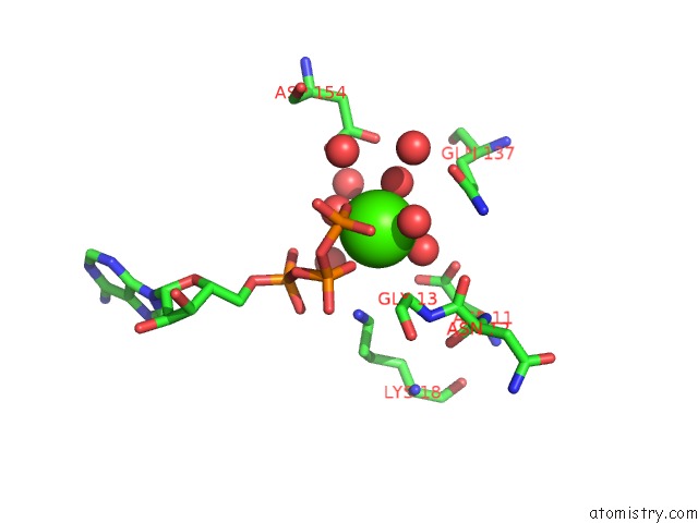
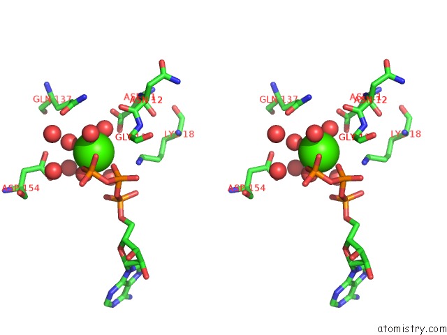
Calcium binding site 1 out of 3 in 3a5n
Go back to
Calcium binding site 1 out
of 3 in the Crystal Structure of A Dictyostelium P109A CA2+-Actin in Complex with Human Gelsolin Segment 1
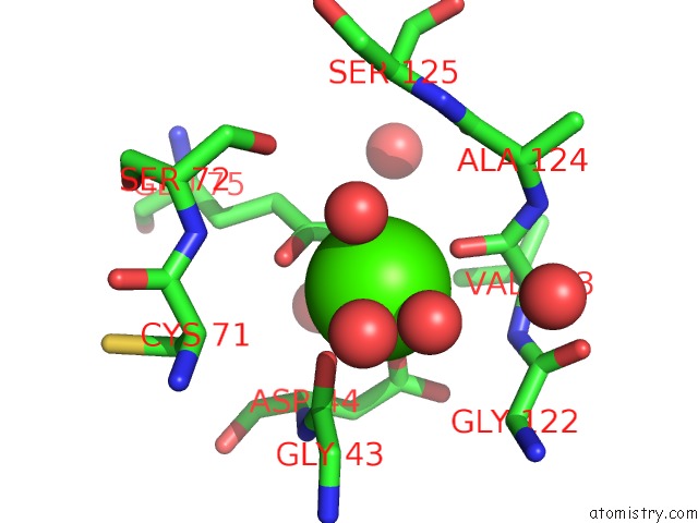
Mono view
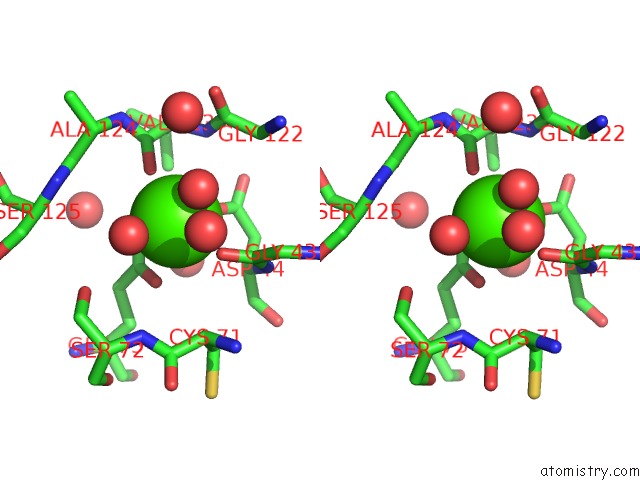
Stereo pair view
Mono view
Stereo pair view
A full contact list of Calcium with other atoms in the Ca binding
site number 1 of Crystal Structure of A Dictyostelium P109A CA2+-Actin in Complex with Human Gelsolin Segment 1 within 5.0Å range:
|
Calcium binding site 2 out of 3 in 3a5n
Go back to
Calcium binding site 2 out
of 3 in the Crystal Structure of A Dictyostelium P109A CA2+-Actin in Complex with Human Gelsolin Segment 1
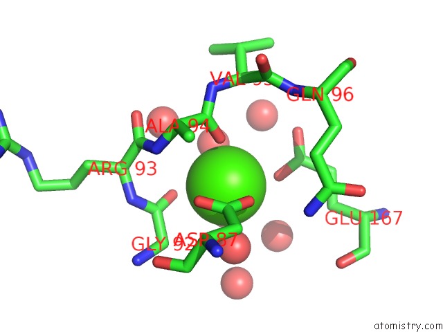
Mono view
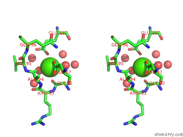
Stereo pair view
Mono view
Stereo pair view
A full contact list of Calcium with other atoms in the Ca binding
site number 2 of Crystal Structure of A Dictyostelium P109A CA2+-Actin in Complex with Human Gelsolin Segment 1 within 5.0Å range:
|
Calcium binding site 3 out of 3 in 3a5n
Go back to
Calcium binding site 3 out
of 3 in the Crystal Structure of A Dictyostelium P109A CA2+-Actin in Complex with Human Gelsolin Segment 1
Mono view
Stereo pair view
Mono view
Stereo pair view
A full contact list of Calcium with other atoms in the Ca binding
site number 3 of Crystal Structure of A Dictyostelium P109A CA2+-Actin in Complex with Human Gelsolin Segment 1 within 5.0Å range:
|
Reference:
K.Murakami,
T.Yasunaga,
T.Q.P.Noguchi,
Y.Gomibuchi,
K.X.Ngo,
T.Q.P.Uyeda,
T.Wakabayashi.
Structural Basis For Actin Assembly, Activation of Atp Hydrolysis, and Delayed Phosphate Release Cell(Cambridge,Mass.) V. 143 275 2010.
ISSN: ISSN 0092-8674
PubMed: 20946985
DOI: 10.1016/J.CELL.2010.09.034
Page generated: Tue Jul 8 10:24:45 2025
ISSN: ISSN 0092-8674
PubMed: 20946985
DOI: 10.1016/J.CELL.2010.09.034
Last articles
Fe in 2YXOFe in 2YRS
Fe in 2YXC
Fe in 2YNM
Fe in 2YVJ
Fe in 2YP1
Fe in 2YU2
Fe in 2YU1
Fe in 2YQB
Fe in 2YOO