Calcium »
PDB 3a4u-3aas »
3a9h »
Calcium in PDB 3a9h: Crystal Structure of Pqq-Dependent Sugar Dehydrogenase Holo-Form
Enzymatic activity of Crystal Structure of Pqq-Dependent Sugar Dehydrogenase Holo-Form
All present enzymatic activity of Crystal Structure of Pqq-Dependent Sugar Dehydrogenase Holo-Form:
1.1.5.2;
1.1.5.2;
Protein crystallography data
The structure of Crystal Structure of Pqq-Dependent Sugar Dehydrogenase Holo-Form, PDB code: 3a9h
was solved by
H.Sakuraba,
K.Yokono,
K.Yoneda,
T.Ohshima,
with X-Ray Crystallography technique. A brief refinement statistics is given in the table below:
| Resolution Low / High (Å) | 44.34 / 2.50 |
| Space group | I 41 2 2 |
| Cell size a, b, c (Å), α, β, γ (°) | 177.344, 177.344, 89.743, 90.00, 90.00, 90.00 |
| R / Rfree (%) | 20.1 / 23.1 |
Calcium Binding Sites:
The binding sites of Calcium atom in the Crystal Structure of Pqq-Dependent Sugar Dehydrogenase Holo-Form
(pdb code 3a9h). This binding sites where shown within
5.0 Angstroms radius around Calcium atom.
In total only one binding site of Calcium was determined in the Crystal Structure of Pqq-Dependent Sugar Dehydrogenase Holo-Form, PDB code: 3a9h:
In total only one binding site of Calcium was determined in the Crystal Structure of Pqq-Dependent Sugar Dehydrogenase Holo-Form, PDB code: 3a9h:
Calcium binding site 1 out of 1 in 3a9h
Go back to
Calcium binding site 1 out
of 1 in the Crystal Structure of Pqq-Dependent Sugar Dehydrogenase Holo-Form
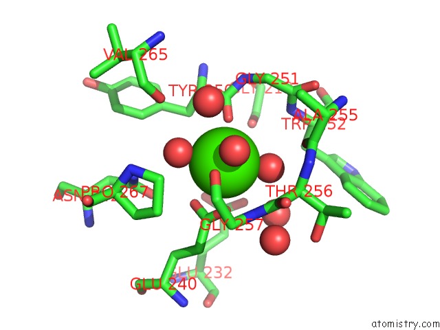
Mono view
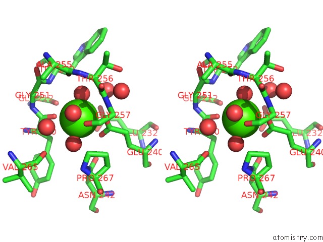
Stereo pair view
Mono view
Stereo pair view
A full contact list of Calcium with other atoms in the Ca binding
site number 1 of Crystal Structure of Pqq-Dependent Sugar Dehydrogenase Holo-Form within 5.0Å range:
|
Reference:
H.Sakuraba,
K.Yokono,
K.Yoneda,
A.Watanabe,
Y.Asada,
T.Satomura,
T.Yabutani,
J.Motonaka,
T.Ohshima.
Catalytic Properties and Crystal Structure of Quinoprotein Aldose Sugar Dehydrogenase From Hyperthermophilic Archaeon Pyrobaculum Aerophilum Arch.Biochem.Biophys. V. 502 81 2010.
ISSN: ISSN 0003-9861
PubMed: 20692227
DOI: 10.1016/J.ABB.2010.08.002
Page generated: Sat Jul 13 07:18:56 2024
ISSN: ISSN 0003-9861
PubMed: 20692227
DOI: 10.1016/J.ABB.2010.08.002
Last articles
Zn in 9J0NZn in 9J0O
Zn in 9J0P
Zn in 9FJX
Zn in 9EKB
Zn in 9C0F
Zn in 9CAH
Zn in 9CH0
Zn in 9CH3
Zn in 9CH1