Calcium »
PDB 3aau-3ati »
3ach »
Calcium in PDB 3ach: Crystal Structure of Carbohydrate-Binding Module Family 28 From Clostridium Josui CEL5A in Complex with Cellotetraose
Enzymatic activity of Crystal Structure of Carbohydrate-Binding Module Family 28 From Clostridium Josui CEL5A in Complex with Cellotetraose
All present enzymatic activity of Crystal Structure of Carbohydrate-Binding Module Family 28 From Clostridium Josui CEL5A in Complex with Cellotetraose:
3.2.1.4;
3.2.1.4;
Protein crystallography data
The structure of Crystal Structure of Carbohydrate-Binding Module Family 28 From Clostridium Josui CEL5A in Complex with Cellotetraose, PDB code: 3ach
was solved by
K.Tsukimoto,
R.Takada,
Y.Araki,
K.Suzuki,
S.Karita,
T.Wakagi,
H.Shoun,
T.Watanabe,
S.Fushinobu,
with X-Ray Crystallography technique. A brief refinement statistics is given in the table below:
| Resolution Low / High (Å) | 27.34 / 1.40 |
| Space group | P 21 21 21 |
| Cell size a, b, c (Å), α, β, γ (°) | 39.576, 63.900, 75.619, 90.00, 90.00, 90.00 |
| R / Rfree (%) | 16.2 / 19.8 |
Calcium Binding Sites:
The binding sites of Calcium atom in the Crystal Structure of Carbohydrate-Binding Module Family 28 From Clostridium Josui CEL5A in Complex with Cellotetraose
(pdb code 3ach). This binding sites where shown within
5.0 Angstroms radius around Calcium atom.
In total only one binding site of Calcium was determined in the Crystal Structure of Carbohydrate-Binding Module Family 28 From Clostridium Josui CEL5A in Complex with Cellotetraose, PDB code: 3ach:
In total only one binding site of Calcium was determined in the Crystal Structure of Carbohydrate-Binding Module Family 28 From Clostridium Josui CEL5A in Complex with Cellotetraose, PDB code: 3ach:
Calcium binding site 1 out of 1 in 3ach
Go back to
Calcium binding site 1 out
of 1 in the Crystal Structure of Carbohydrate-Binding Module Family 28 From Clostridium Josui CEL5A in Complex with Cellotetraose
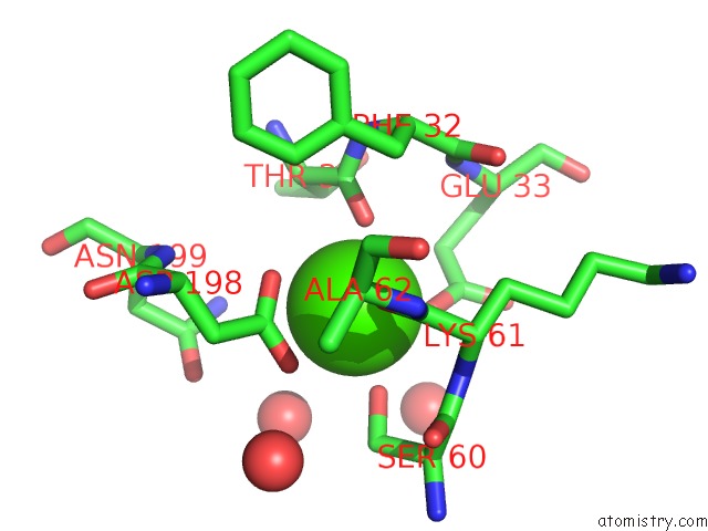
Mono view
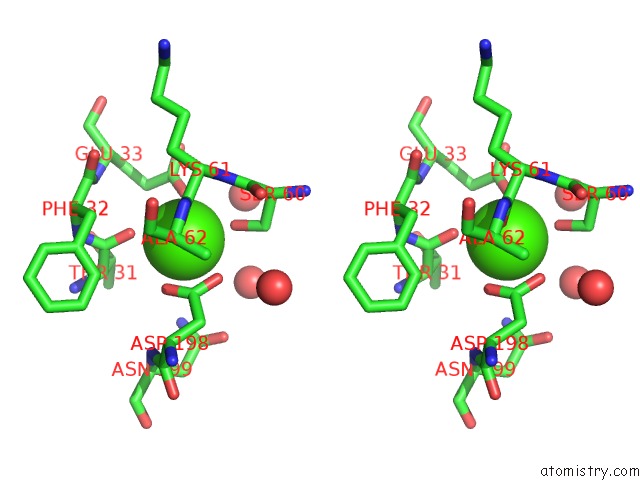
Stereo pair view
Mono view
Stereo pair view
A full contact list of Calcium with other atoms in the Ca binding
site number 1 of Crystal Structure of Carbohydrate-Binding Module Family 28 From Clostridium Josui CEL5A in Complex with Cellotetraose within 5.0Å range:
|
Reference:
K.Tsukimoto,
R.Takada,
Y.Araki,
K.Suzuki,
S.Karita,
T.Wakagi,
H.Shoun,
T.Watanabe,
S.Fushinobu.
Recognition of Cellooligosaccharides By A Family 28 Carbohydrate-Binding Module. Febs Lett. V. 584 1205 2010.
ISSN: ISSN 0014-5793
PubMed: 20159017
DOI: 10.1016/J.FEBSLET.2010.02.027
Page generated: Sat Jul 13 07:44:58 2024
ISSN: ISSN 0014-5793
PubMed: 20159017
DOI: 10.1016/J.FEBSLET.2010.02.027
Last articles
Zn in 9MJ5Zn in 9HNW
Zn in 9G0L
Zn in 9FNE
Zn in 9DZN
Zn in 9E0I
Zn in 9D32
Zn in 9DAK
Zn in 8ZXC
Zn in 8ZUF