Calcium »
PDB 3aau-3ati »
3alt »
Calcium in PDB 3alt: Crystal Structure of Cel-IV Complexed with Melibiose
Protein crystallography data
The structure of Crystal Structure of Cel-IV Complexed with Melibiose, PDB code: 3alt
was solved by
T.Hatakeyama,
T.Hozawa,
K.Ishii,
T.Kamiya,
S.Goda,
M.Kusunoki,
H.Unno,
with X-Ray Crystallography technique. A brief refinement statistics is given in the table below:
| Resolution Low / High (Å) | 38.06 / 2.50 |
| Space group | P 1 21 1 |
| Cell size a, b, c (Å), α, β, γ (°) | 43.988, 78.504, 102.898, 90.00, 98.41, 90.00 |
| R / Rfree (%) | 22.9 / 27.9 |
Calcium Binding Sites:
The binding sites of Calcium atom in the Crystal Structure of Cel-IV Complexed with Melibiose
(pdb code 3alt). This binding sites where shown within
5.0 Angstroms radius around Calcium atom.
In total 4 binding sites of Calcium where determined in the Crystal Structure of Cel-IV Complexed with Melibiose, PDB code: 3alt:
Jump to Calcium binding site number: 1; 2; 3; 4;
In total 4 binding sites of Calcium where determined in the Crystal Structure of Cel-IV Complexed with Melibiose, PDB code: 3alt:
Jump to Calcium binding site number: 1; 2; 3; 4;
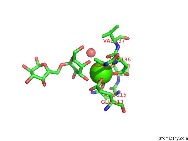
Calcium binding site 1 out of 4 in 3alt
Go back to
Calcium binding site 1 out
of 4 in the Crystal Structure of Cel-IV Complexed with Melibiose
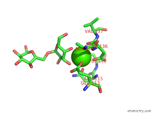
Mono view
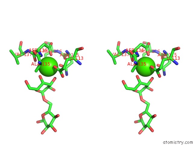
Stereo pair view
Mono view
Stereo pair view
A full contact list of Calcium with other atoms in the Ca binding
site number 1 of Crystal Structure of Cel-IV Complexed with Melibiose within 5.0Å range:
|
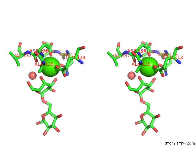
Calcium binding site 2 out of 4 in 3alt
Go back to
Calcium binding site 2 out
of 4 in the Crystal Structure of Cel-IV Complexed with Melibiose
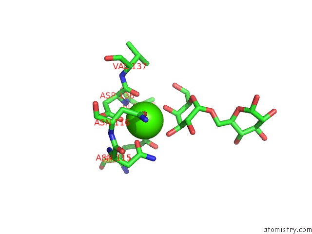
Mono view
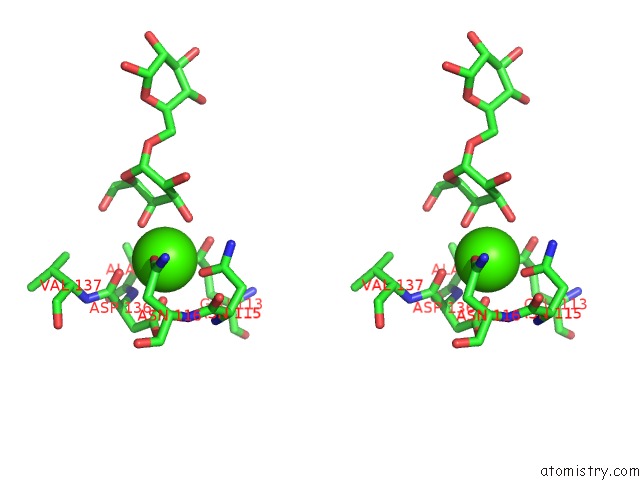
Stereo pair view
Mono view
Stereo pair view
A full contact list of Calcium with other atoms in the Ca binding
site number 2 of Crystal Structure of Cel-IV Complexed with Melibiose within 5.0Å range:
|
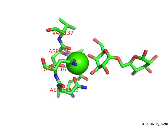
Calcium binding site 3 out of 4 in 3alt
Go back to
Calcium binding site 3 out
of 4 in the Crystal Structure of Cel-IV Complexed with Melibiose
Mono view
Stereo pair view
Mono view
Stereo pair view
A full contact list of Calcium with other atoms in the Ca binding
site number 3 of Crystal Structure of Cel-IV Complexed with Melibiose within 5.0Å range:
|
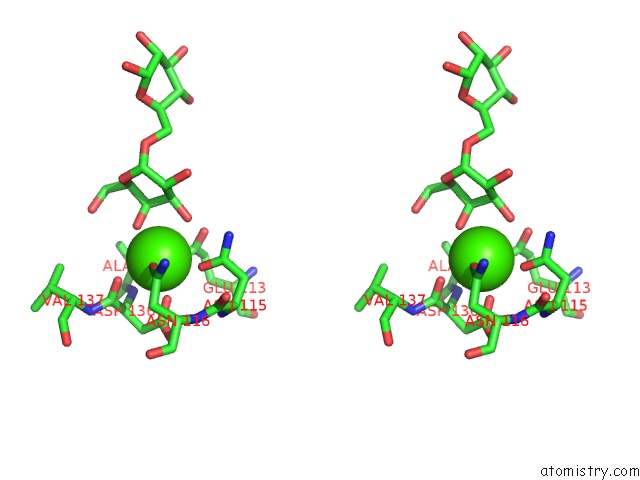
Calcium binding site 4 out of 4 in 3alt
Go back to
Calcium binding site 4 out
of 4 in the Crystal Structure of Cel-IV Complexed with Melibiose
Mono view
Stereo pair view
Mono view
Stereo pair view
A full contact list of Calcium with other atoms in the Ca binding
site number 4 of Crystal Structure of Cel-IV Complexed with Melibiose within 5.0Å range:
|
Reference:
T.Hatakeyama,
T.Kamiya,
M.Kusunoki,
S.Nakamura-Tsuruta,
J.Hirabayashi,
S.Goda,
H.Unno.
Galactose Recognition By A Tetrameric C-Type Lectin, Cel-IV, Containing the Epn Carbohydrate Recognition Motif J.Biol.Chem. V. 286 10305 2011.
ISSN: ISSN 0021-9258
PubMed: 21247895
DOI: 10.1074/JBC.M110.200576
Page generated: Sat Jul 13 07:52:25 2024
ISSN: ISSN 0021-9258
PubMed: 21247895
DOI: 10.1074/JBC.M110.200576
Last articles
Zn in 9MJ5Zn in 9HNW
Zn in 9G0L
Zn in 9FNE
Zn in 9DZN
Zn in 9E0I
Zn in 9D32
Zn in 9DAK
Zn in 8ZXC
Zn in 8ZUF