Calcium »
PDB 3atj-3b1t »
3axh »
Calcium in PDB 3axh: Crystal Structure of Isomaltase in Complex with Isomaltose
Enzymatic activity of Crystal Structure of Isomaltase in Complex with Isomaltose
All present enzymatic activity of Crystal Structure of Isomaltase in Complex with Isomaltose:
3.2.1.10;
3.2.1.10;
Protein crystallography data
The structure of Crystal Structure of Isomaltase in Complex with Isomaltose, PDB code: 3axh
was solved by
K.Yamamoto,
H.Miyake,
M.Kusunoki,
S.Osaki,
with X-Ray Crystallography technique. A brief refinement statistics is given in the table below:
| Resolution Low / High (Å) | 24.38 / 1.80 |
| Space group | C 1 2 1 |
| Cell size a, b, c (Å), α, β, γ (°) | 94.892, 114.736, 61.496, 90.00, 90.99, 90.00 |
| R / Rfree (%) | 16.8 / 19.3 |
Calcium Binding Sites:
The binding sites of Calcium atom in the Crystal Structure of Isomaltase in Complex with Isomaltose
(pdb code 3axh). This binding sites where shown within
5.0 Angstroms radius around Calcium atom.
In total only one binding site of Calcium was determined in the Crystal Structure of Isomaltase in Complex with Isomaltose, PDB code: 3axh:
In total only one binding site of Calcium was determined in the Crystal Structure of Isomaltase in Complex with Isomaltose, PDB code: 3axh:
Calcium binding site 1 out of 1 in 3axh
Go back to
Calcium binding site 1 out
of 1 in the Crystal Structure of Isomaltase in Complex with Isomaltose
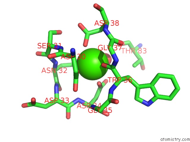
Mono view
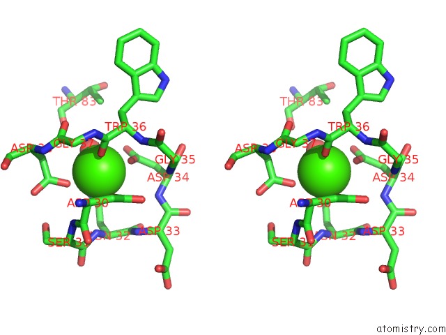
Stereo pair view
Mono view
Stereo pair view
A full contact list of Calcium with other atoms in the Ca binding
site number 1 of Crystal Structure of Isomaltase in Complex with Isomaltose within 5.0Å range:
|
Reference:
K.Yamamoto,
H.Miyake,
M.Kusunoki,
S.Osaki.
Steric Hindrance By 2 Amino Acid Residues Determines the Substrate Specificity of Isomaltase From Saccharomyces Cerevisiae J.Biosci.Bioeng. V. 112 545 2011.
ISSN: ISSN 1389-1723
PubMed: 21925939
DOI: 10.1016/J.JBIOSC.2011.08.016
Page generated: Sat Jul 13 08:02:40 2024
ISSN: ISSN 1389-1723
PubMed: 21925939
DOI: 10.1016/J.JBIOSC.2011.08.016
Last articles
Zn in 9J0NZn in 9J0O
Zn in 9J0P
Zn in 9FJX
Zn in 9EKB
Zn in 9C0F
Zn in 9CAH
Zn in 9CH0
Zn in 9CH3
Zn in 9CH1