Calcium »
PDB 3atj-3b1t »
3ayf »
Calcium in PDB 3ayf: Crystal Structure of Nitric Oxide Reductase
Protein crystallography data
The structure of Crystal Structure of Nitric Oxide Reductase, PDB code: 3ayf
was solved by
Y.Matsumoto,
T.Tosha,
A.V.Pisliakov,
T.Hino,
H.Sugimoti,
S.Nagano,
Y.Sugita,
Y.Shiro,
with X-Ray Crystallography technique. A brief refinement statistics is given in the table below:
| Resolution Low / High (Å) | 19.78 / 2.50 |
| Space group | C 2 2 21 |
| Cell size a, b, c (Å), α, β, γ (°) | 110.409, 149.648, 151.108, 90.00, 90.00, 90.00 |
| R / Rfree (%) | 24.4 / 28.2 |
Other elements in 3ayf:
The structure of Crystal Structure of Nitric Oxide Reductase also contains other interesting chemical elements:
| Iron | (Fe) | 2 atoms |
| Zinc | (Zn) | 1 atom |
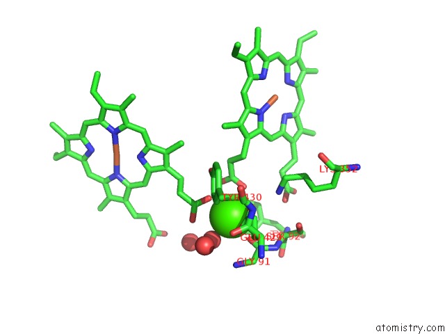
Calcium Binding Sites:
The binding sites of Calcium atom in the Crystal Structure of Nitric Oxide Reductase
(pdb code 3ayf). This binding sites where shown within
5.0 Angstroms radius around Calcium atom.
In total only one binding site of Calcium was determined in the Crystal Structure of Nitric Oxide Reductase, PDB code: 3ayf:
In total only one binding site of Calcium was determined in the Crystal Structure of Nitric Oxide Reductase, PDB code: 3ayf:
Calcium binding site 1 out of 1 in 3ayf
Go back to
Calcium binding site 1 out
of 1 in the Crystal Structure of Nitric Oxide Reductase
Mono view
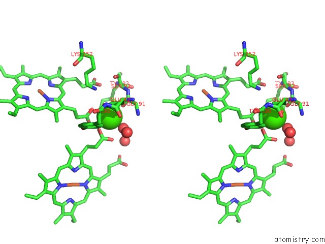
Stereo pair view
Mono view
Stereo pair view
A full contact list of Calcium with other atoms in the Ca binding
site number 1 of Crystal Structure of Nitric Oxide Reductase within 5.0Å range:
|
Reference:
Y.Matsumoto,
T.Tosha,
A.V.Pisliakov,
T.Hino,
H.Sugimoto,
S.Nagano,
Y.Sugita,
Y.Shiro.
Crystal Structure of Quinol-Dependent Nitric Oxide Reductase From Geobacillus Stearothermophilus Nat.Struct.Mol.Biol. V. 19 238 2012.
ISSN: ISSN 1545-9993
PubMed: 22266822
DOI: 10.1038/NSMB.2213
Page generated: Sat Jul 13 08:03:25 2024
ISSN: ISSN 1545-9993
PubMed: 22266822
DOI: 10.1038/NSMB.2213
Last articles
Zn in 9JYWZn in 9IR4
Zn in 9IR3
Zn in 9GMX
Zn in 9GMW
Zn in 9JEJ
Zn in 9ERF
Zn in 9ERE
Zn in 9EGV
Zn in 9EGW