Calcium in PDB 3ba0: Crystal Structure of Full-Length Human Mmp-12
Enzymatic activity of Crystal Structure of Full-Length Human Mmp-12
All present enzymatic activity of Crystal Structure of Full-Length Human Mmp-12:
3.4.24.65;
Protein crystallography data
The structure of Crystal Structure of Full-Length Human Mmp-12, PDB code: 3ba0
was solved by
I.Bertini,
V.Calderone,
M.Fragai,
R.Jaiswal,
C.Luchinat,
M.Melikian,
E.Myonas,
D.I.Svergun,
with X-Ray Crystallography technique. A brief refinement statistics is given in the table below:
Resolution Low / High (Å)
|
30.70 /
3.00
|
Space group
|
C 1 2 1
|
Cell size a, b, c (Å), α, β, γ (°)
|
135.037,
60.148,
59.611,
90.00,
90.74,
90.00
|
R / Rfree (%)
|
23.6 /
31.9
|
Other elements in 3ba0:
The structure of Crystal Structure of Full-Length Human Mmp-12 also contains other interesting chemical elements:
Calcium Binding Sites:
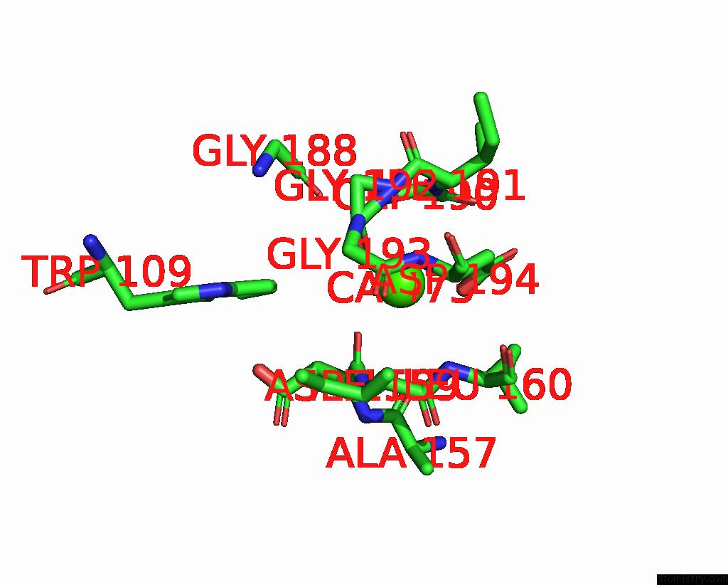
The binding sites of Calcium atom in the Crystal Structure of Full-Length Human Mmp-12
(pdb code 3ba0). This binding sites where shown within
5.0 Angstroms radius around Calcium atom.
In total 4 binding sites of Calcium where determined in the
Crystal Structure of Full-Length Human Mmp-12, PDB code: 3ba0:
Jump to Calcium binding site number:
1;
2;
3;
4;
Calcium binding site 1 out
of 4 in 3ba0
Go back to
Calcium Binding Sites List in 3ba0
Calcium binding site 1 out
of 4 in the Crystal Structure of Full-Length Human Mmp-12
Mono view
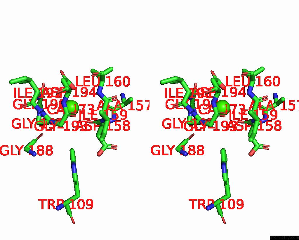
Stereo pair view
|
A full contact list of Calcium with other atoms in the Ca binding
site number 1 of Crystal Structure of Full-Length Human Mmp-12 within 5.0Å range:
probe
|
atom
|
residue
|
distance (Å)
|
B
|
Occ
|
A:Ca473
b:2.0
occ:1.00
|
O
|
A:GLY192
|
2.2
|
2.4
|
1.0
|
O
|
A:ASP158
|
2.6
|
6.9
|
1.0
|
OD2
|
A:ASP194
|
3.1
|
2.7
|
1.0
|
C
|
A:GLY192
|
3.5
|
6.9
|
1.0
|
C
|
A:ASP158
|
3.6
|
6.6
|
1.0
|
N
|
A:GLY192
|
3.8
|
10.4
|
1.0
|
N
|
A:ILE191
|
4.0
|
7.9
|
1.0
|
CG
|
A:ASP194
|
4.1
|
2.0
|
1.0
|
CA
|
A:ASP158
|
4.2
|
9.4
|
1.0
|
CA
|
A:ILE191
|
4.2
|
9.8
|
1.0
|
C
|
A:GLY190
|
4.3
|
8.7
|
1.0
|
CA
|
A:GLY192
|
4.4
|
7.9
|
1.0
|
N
|
A:GLY193
|
4.4
|
4.8
|
1.0
|
N
|
A:ILE159
|
4.4
|
6.2
|
1.0
|
N
|
A:ASP194
|
4.4
|
2.0
|
1.0
|
C
|
A:ILE191
|
4.5
|
11.8
|
1.0
|
OD1
|
A:ASP194
|
4.6
|
2.0
|
1.0
|
O
|
A:GLY188
|
4.6
|
5.7
|
1.0
|
N
|
A:GLY190
|
4.6
|
6.5
|
1.0
|
O
|
A:ALA157
|
4.6
|
11.3
|
1.0
|
CA
|
A:ILE159
|
4.6
|
4.5
|
1.0
|
CB
|
A:ASP158
|
4.7
|
10.9
|
1.0
|
N
|
A:LEU160
|
4.8
|
2.0
|
1.0
|
O
|
A:GLY190
|
4.9
|
12.6
|
1.0
|
CH2
|
A:TRP109
|
4.9
|
2.0
|
1.0
|
C
|
A:GLY193
|
4.9
|
2.0
|
1.0
|
CA
|
A:GLY190
|
4.9
|
8.4
|
1.0
|
CA
|
A:GLY193
|
4.9
|
2.0
|
1.0
|
CD1
|
A:LEU160
|
5.0
|
2.0
|
1.0
|
C
|
A:ILE159
|
5.0
|
2.8
|
1.0
|
|
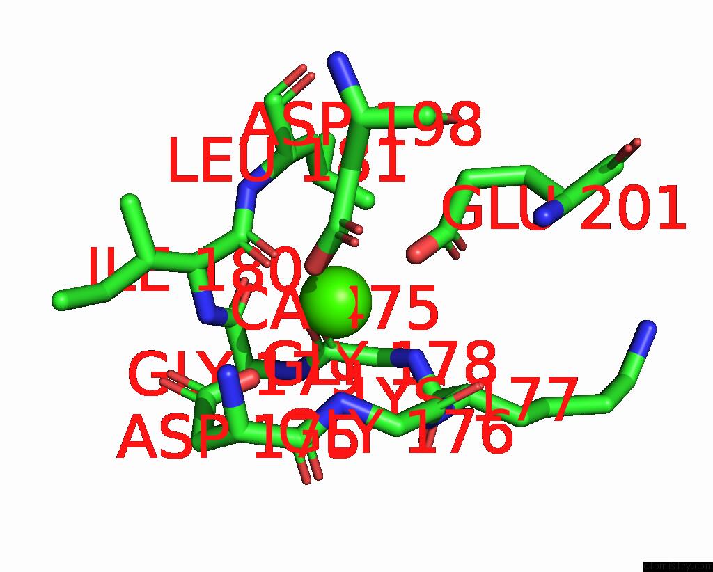
Calcium binding site 2 out
of 4 in 3ba0
Go back to
Calcium Binding Sites List in 3ba0
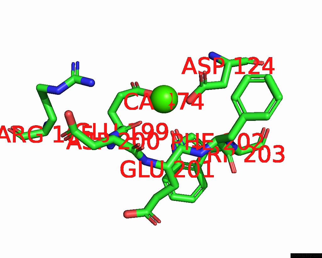
Calcium binding site 2 out
of 4 in the Crystal Structure of Full-Length Human Mmp-12
Mono view
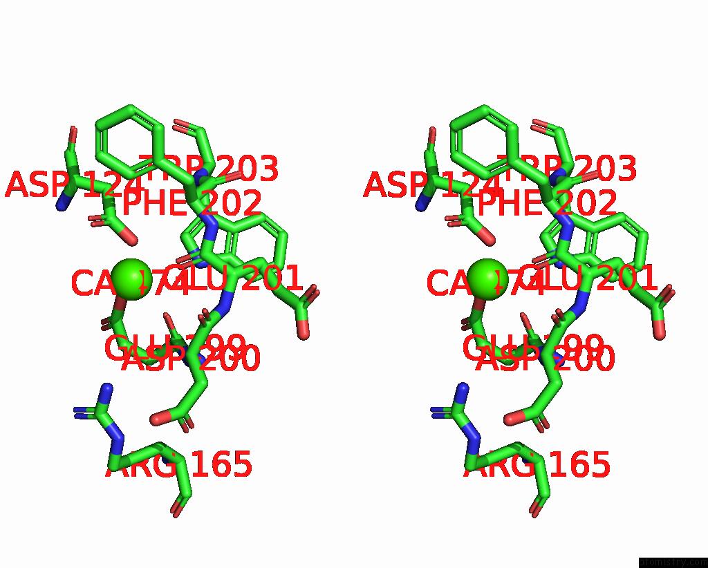
Stereo pair view
|
A full contact list of Calcium with other atoms in the Ca binding
site number 2 of Crystal Structure of Full-Length Human Mmp-12 within 5.0Å range:
probe
|
atom
|
residue
|
distance (Å)
|
B
|
Occ
|
A:Ca474
b:2.0
occ:1.00
|
O
|
A:GLU201
|
2.2
|
2.0
|
1.0
|
O
|
A:GLU199
|
2.3
|
4.0
|
1.0
|
OD2
|
A:ASP124
|
2.5
|
2.0
|
1.0
|
OE2
|
A:GLU199
|
3.0
|
2.0
|
1.0
|
OD1
|
A:ASP124
|
3.1
|
10.9
|
1.0
|
CG
|
A:ASP124
|
3.2
|
7.5
|
1.0
|
C
|
A:GLU201
|
3.4
|
3.2
|
1.0
|
C
|
A:GLU199
|
3.5
|
2.0
|
1.0
|
CG
|
A:GLU199
|
3.9
|
2.0
|
1.0
|
CD
|
A:GLU199
|
4.0
|
2.0
|
1.0
|
C
|
A:ASP200
|
4.0
|
6.5
|
1.0
|
N
|
A:GLU201
|
4.2
|
6.8
|
1.0
|
CA
|
A:ASP200
|
4.2
|
5.2
|
1.0
|
N
|
A:PHE202
|
4.3
|
3.6
|
1.0
|
O
|
A:ASP200
|
4.3
|
7.7
|
1.0
|
CA
|
A:PHE202
|
4.3
|
2.0
|
1.0
|
N
|
A:ASP200
|
4.4
|
2.7
|
1.0
|
CA
|
A:GLU199
|
4.4
|
2.0
|
1.0
|
CA
|
A:GLU201
|
4.4
|
4.0
|
1.0
|
CD1
|
A:PHE202
|
4.6
|
11.4
|
1.0
|
CB
|
A:GLU199
|
4.7
|
2.0
|
1.0
|
CB
|
A:ASP124
|
4.8
|
2.0
|
1.0
|
NH2
|
A:ARG165
|
4.8
|
2.0
|
1.0
|
CD1
|
A:TRP203
|
5.0
|
2.0
|
1.0
|
|
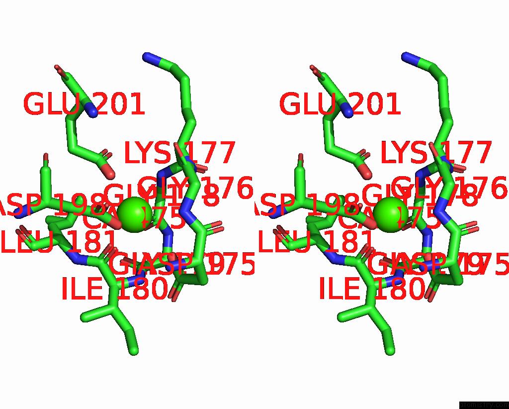
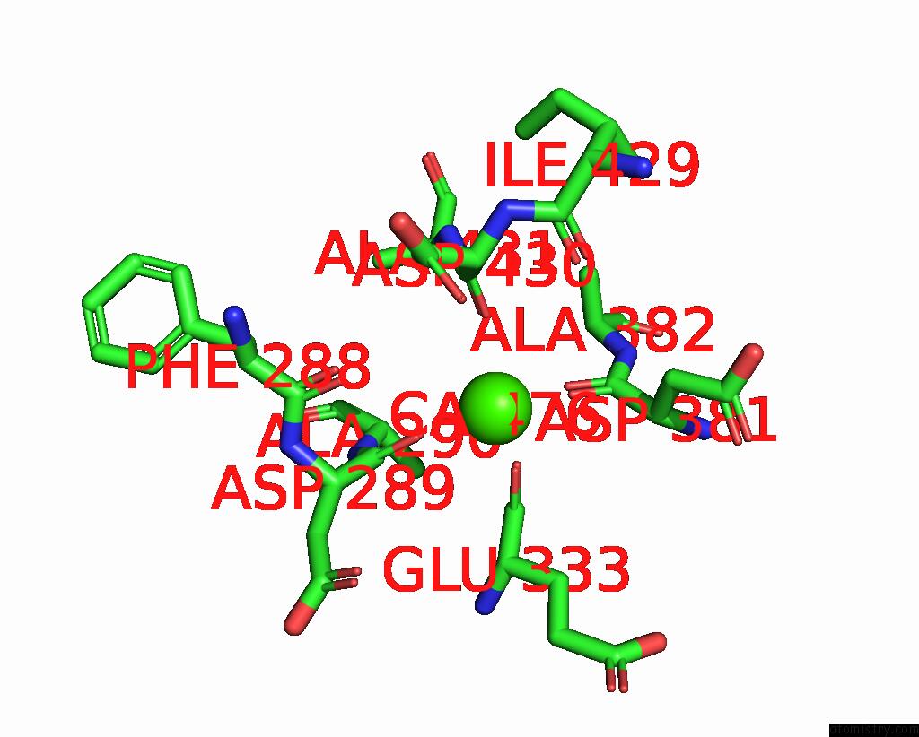
Calcium binding site 3 out
of 4 in 3ba0
Go back to
Calcium Binding Sites List in 3ba0
Calcium binding site 3 out
of 4 in the Crystal Structure of Full-Length Human Mmp-12
Mono view
Stereo pair view
|
A full contact list of Calcium with other atoms in the Ca binding
site number 3 of Crystal Structure of Full-Length Human Mmp-12 within 5.0Å range:
probe
|
atom
|
residue
|
distance (Å)
|
B
|
Occ
|
A:Ca475
b:2.0
occ:1.00
|
OD2
|
A:ASP198
|
1.7
|
3.0
|
1.0
|
O
|
A:ILE180
|
2.2
|
2.0
|
1.0
|
OE2
|
A:GLU201
|
2.5
|
2.0
|
1.0
|
O
|
A:GLY178
|
2.6
|
2.0
|
1.0
|
CG
|
A:ASP198
|
2.8
|
4.8
|
1.0
|
OD2
|
A:ASP175
|
2.9
|
2.6
|
1.0
|
N
|
A:GLY176
|
3.2
|
2.0
|
1.0
|
C
|
A:ILE180
|
3.5
|
2.0
|
1.0
|
CB
|
A:ASP198
|
3.6
|
2.2
|
1.0
|
OD1
|
A:ASP198
|
3.7
|
2.0
|
1.0
|
N
|
A:GLY178
|
3.7
|
2.8
|
1.0
|
CD
|
A:GLU201
|
3.7
|
2.0
|
1.0
|
C
|
A:GLY178
|
3.7
|
2.0
|
1.0
|
C
|
A:GLY176
|
3.7
|
2.0
|
1.0
|
N
|
A:LYS177
|
3.7
|
2.0
|
1.0
|
C
|
A:ASP175
|
3.9
|
2.0
|
1.0
|
CA
|
A:GLY176
|
3.9
|
2.0
|
1.0
|
CG
|
A:ASP175
|
4.0
|
2.0
|
1.0
|
CA
|
A:LYS177
|
4.2
|
2.0
|
1.0
|
N
|
A:ASP175
|
4.2
|
2.0
|
1.0
|
O
|
A:GLY176
|
4.2
|
2.0
|
1.0
|
CA
|
A:GLY178
|
4.3
|
2.5
|
1.0
|
N
|
A:ILE180
|
4.3
|
2.0
|
1.0
|
C
|
A:LYS177
|
4.3
|
2.2
|
1.0
|
OE1
|
A:GLU201
|
4.3
|
4.5
|
1.0
|
N
|
A:LEU181
|
4.4
|
2.0
|
1.0
|
CA
|
A:ILE180
|
4.4
|
2.0
|
1.0
|
CA
|
A:LEU181
|
4.5
|
2.0
|
1.0
|
CA
|
A:ASP175
|
4.5
|
2.0
|
1.0
|
O
|
A:ASP175
|
4.5
|
2.0
|
1.0
|
OD1
|
A:ASP175
|
4.6
|
2.0
|
1.0
|
C
|
A:GLY179
|
4.7
|
2.0
|
1.0
|
CG
|
A:GLU201
|
4.7
|
4.9
|
1.0
|
N
|
A:GLY179
|
4.8
|
2.0
|
1.0
|
CB
|
A:ASP175
|
4.9
|
2.0
|
1.0
|
O
|
A:GLY179
|
4.9
|
2.6
|
1.0
|
CD2
|
A:LEU181
|
5.0
|
2.0
|
1.0
|
|
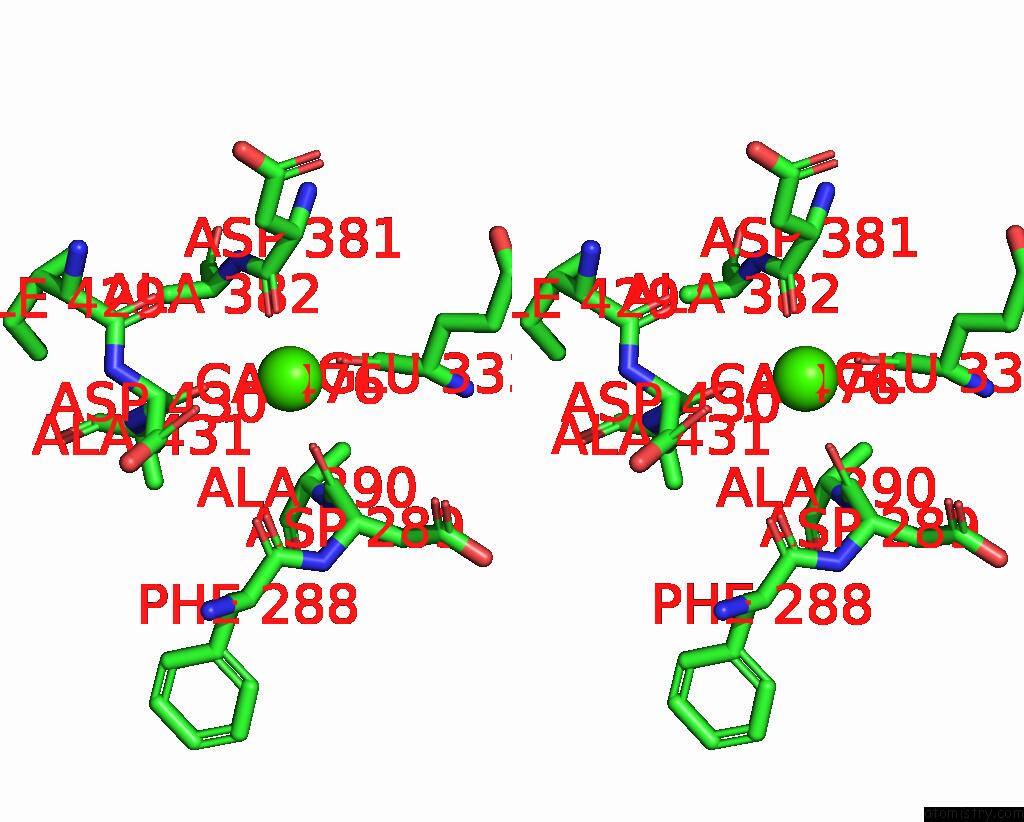
Calcium binding site 4 out
of 4 in 3ba0
Go back to
Calcium Binding Sites List in 3ba0
Calcium binding site 4 out
of 4 in the Crystal Structure of Full-Length Human Mmp-12
Mono view
Stereo pair view
|
A full contact list of Calcium with other atoms in the Ca binding
site number 4 of Crystal Structure of Full-Length Human Mmp-12 within 5.0Å range:
probe
|
atom
|
residue
|
distance (Å)
|
B
|
Occ
|
A:Ca476
b:15.3
occ:1.00
|
O
|
A:ASP289
|
2.2
|
2.0
|
1.0
|
O
|
A:ASP381
|
2.6
|
2.0
|
1.0
|
O
|
A:GLU333
|
2.8
|
2.0
|
1.0
|
O
|
A:ASP430
|
2.8
|
2.7
|
1.0
|
C
|
A:ASP289
|
3.5
|
2.0
|
1.0
|
C
|
A:ASP430
|
3.6
|
2.0
|
1.0
|
CA
|
A:ASP430
|
3.8
|
2.0
|
1.0
|
C
|
A:ASP381
|
3.8
|
2.0
|
1.0
|
C
|
A:GLU333
|
3.9
|
2.0
|
1.0
|
O
|
A:PHE288
|
4.1
|
2.0
|
1.0
|
O
|
A:ILE429
|
4.3
|
2.5
|
1.0
|
OD1
|
A:ASP430
|
4.3
|
2.4
|
1.0
|
CA
|
A:ASP381
|
4.4
|
2.0
|
1.0
|
N
|
A:ALA290
|
4.4
|
2.0
|
1.0
|
CA
|
A:ASP289
|
4.4
|
2.0
|
1.0
|
CB
|
A:ASP430
|
4.4
|
2.0
|
1.0
|
CA
|
A:GLU333
|
4.5
|
2.0
|
1.0
|
CB
|
A:ASP381
|
4.5
|
2.0
|
1.0
|
CA
|
A:ALA290
|
4.7
|
2.0
|
1.0
|
N
|
A:ALA382
|
4.9
|
2.0
|
1.0
|
N
|
A:ALA431
|
4.9
|
2.0
|
1.0
|
CB
|
A:GLU333
|
4.9
|
2.7
|
1.0
|
CG
|
A:ASP430
|
4.9
|
2.0
|
1.0
|
C
|
A:PHE288
|
5.0
|
2.0
|
1.0
|
N
|
A:ASP430
|
5.0
|
2.0
|
1.0
|
|
Reference:
I.Bertini,
V.Calderone,
M.Fragai,
R.Jaiswal,
C.Luchinat,
M.Melikian,
E.Mylonas,
D.I.Svergun.
Evidence of Reciprocal Reorientation of the Catalytic and Hemopexin-Like Domains of Full-Length Mmp-12. J.Am.Chem.Soc. V. 130 7011 2008.
ISSN: ISSN 0002-7863
PubMed: 18465858
DOI: 10.1021/JA710491Y
Page generated: Sat Jul 13 08:11:55 2024
|