Calcium »
PDB 3bje-3bxk »
3btk »
Calcium in PDB 3btk: The Crystal Structures of the Complexes Between Bovine Beta- Trypsin and Ten P1 Variants of Bpti
Enzymatic activity of The Crystal Structures of the Complexes Between Bovine Beta- Trypsin and Ten P1 Variants of Bpti
All present enzymatic activity of The Crystal Structures of the Complexes Between Bovine Beta- Trypsin and Ten P1 Variants of Bpti:
3.4.21.4;
3.4.21.4;
Protein crystallography data
The structure of The Crystal Structures of the Complexes Between Bovine Beta- Trypsin and Ten P1 Variants of Bpti, PDB code: 3btk
was solved by
R.Helland,
J.Otlewski,
O.Sundheim,
M.Dadlez,
A.O.Smalas,
with X-Ray Crystallography technique. A brief refinement statistics is given in the table below:
| Resolution Low / High (Å) | 8.00 / 1.85 |
| Space group | I 2 2 2 |
| Cell size a, b, c (Å), α, β, γ (°) | 75.370, 84.570, 122.810, 90.00, 90.00, 90.00 |
| R / Rfree (%) | 20.2 / 23 |
Calcium Binding Sites:
The binding sites of Calcium atom in the The Crystal Structures of the Complexes Between Bovine Beta- Trypsin and Ten P1 Variants of Bpti
(pdb code 3btk). This binding sites where shown within
5.0 Angstroms radius around Calcium atom.
In total only one binding site of Calcium was determined in the The Crystal Structures of the Complexes Between Bovine Beta- Trypsin and Ten P1 Variants of Bpti, PDB code: 3btk:
In total only one binding site of Calcium was determined in the The Crystal Structures of the Complexes Between Bovine Beta- Trypsin and Ten P1 Variants of Bpti, PDB code: 3btk:
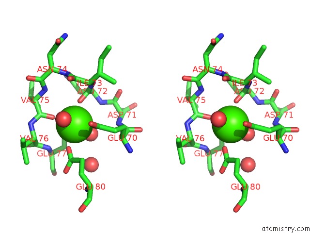
Calcium binding site 1 out of 1 in 3btk
Go back to
Calcium binding site 1 out
of 1 in the The Crystal Structures of the Complexes Between Bovine Beta- Trypsin and Ten P1 Variants of Bpti
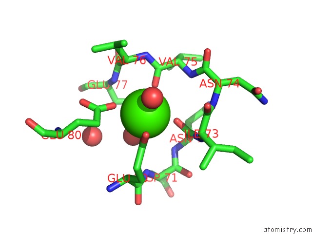
Mono view
Stereo pair view
Mono view
Stereo pair view
A full contact list of Calcium with other atoms in the Ca binding
site number 1 of The Crystal Structures of the Complexes Between Bovine Beta- Trypsin and Ten P1 Variants of Bpti within 5.0Å range:
|
Reference:
R.Helland,
J.Otlewski,
O.Sundheim,
M.Dadlez,
A.O.Smalas.
The Crystal Structures of the Complexes Between Bovine Beta-Trypsin and Ten P1 Variants of Bpti. J.Mol.Biol. V. 287 923 1999.
ISSN: ISSN 0022-2836
PubMed: 10222201
DOI: 10.1006/JMBI.1999.2654
Page generated: Sat Jul 13 08:25:01 2024
ISSN: ISSN 0022-2836
PubMed: 10222201
DOI: 10.1006/JMBI.1999.2654
Last articles
Zn in 9J0NZn in 9J0O
Zn in 9J0P
Zn in 9FJX
Zn in 9EKB
Zn in 9C0F
Zn in 9CAH
Zn in 9CH0
Zn in 9CH3
Zn in 9CH1