Calcium »
PDB 3bje-3bxk »
3bvh »
Calcium in PDB 3bvh: Crystal Structure of Recombinant GAMMAD364A Fibrinogen Fragment D with the Peptide Ligand Gly-Pro-Arg-Pro-Amide
Protein crystallography data
The structure of Crystal Structure of Recombinant GAMMAD364A Fibrinogen Fragment D with the Peptide Ligand Gly-Pro-Arg-Pro-Amide, PDB code: 3bvh
was solved by
S.R.Bowley,
B.K.Merenbloom,
L.Betts,
N.Okumura,
A.Heroux,
O.V.Gorkun,
S.T.Lord,
with X-Ray Crystallography technique. A brief refinement statistics is given in the table below:
| Resolution Low / High (Å) | 50.00 / 2.60 |
| Space group | P 21 21 21 |
| Cell size a, b, c (Å), α, β, γ (°) | 89.844, 94.913, 225.754, 90.00, 90.00, 90.00 |
| R / Rfree (%) | 22 / 26 |
Calcium Binding Sites:
The binding sites of Calcium atom in the Crystal Structure of Recombinant GAMMAD364A Fibrinogen Fragment D with the Peptide Ligand Gly-Pro-Arg-Pro-Amide
(pdb code 3bvh). This binding sites where shown within
5.0 Angstroms radius around Calcium atom.
In total 4 binding sites of Calcium where determined in the Crystal Structure of Recombinant GAMMAD364A Fibrinogen Fragment D with the Peptide Ligand Gly-Pro-Arg-Pro-Amide, PDB code: 3bvh:
Jump to Calcium binding site number: 1; 2; 3; 4;
In total 4 binding sites of Calcium where determined in the Crystal Structure of Recombinant GAMMAD364A Fibrinogen Fragment D with the Peptide Ligand Gly-Pro-Arg-Pro-Amide, PDB code: 3bvh:
Jump to Calcium binding site number: 1; 2; 3; 4;
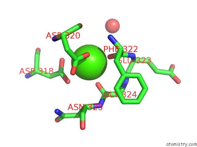
Calcium binding site 1 out of 4 in 3bvh
Go back to
Calcium binding site 1 out
of 4 in the Crystal Structure of Recombinant GAMMAD364A Fibrinogen Fragment D with the Peptide Ligand Gly-Pro-Arg-Pro-Amide
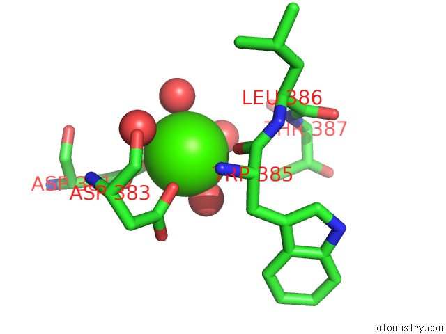
Mono view
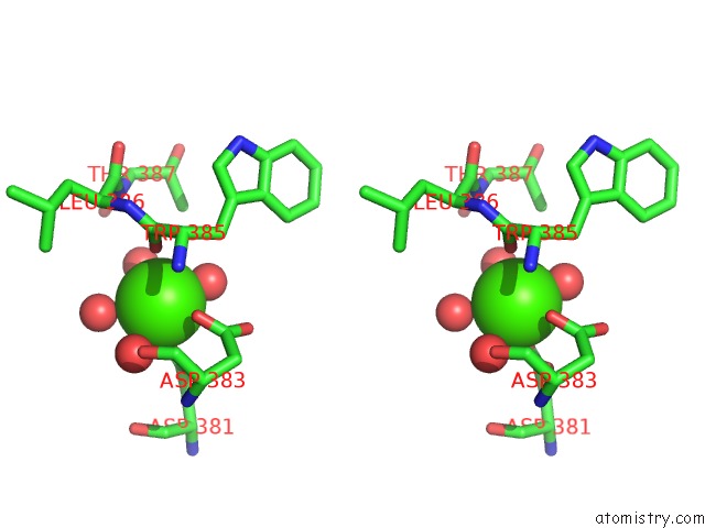
Stereo pair view
Mono view
Stereo pair view
A full contact list of Calcium with other atoms in the Ca binding
site number 1 of Crystal Structure of Recombinant GAMMAD364A Fibrinogen Fragment D with the Peptide Ligand Gly-Pro-Arg-Pro-Amide within 5.0Å range:
|
Calcium binding site 2 out of 4 in 3bvh
Go back to
Calcium binding site 2 out
of 4 in the Crystal Structure of Recombinant GAMMAD364A Fibrinogen Fragment D with the Peptide Ligand Gly-Pro-Arg-Pro-Amide
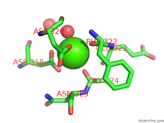
Mono view

Stereo pair view
Mono view
Stereo pair view
A full contact list of Calcium with other atoms in the Ca binding
site number 2 of Crystal Structure of Recombinant GAMMAD364A Fibrinogen Fragment D with the Peptide Ligand Gly-Pro-Arg-Pro-Amide within 5.0Å range:
|
Calcium binding site 3 out of 4 in 3bvh
Go back to
Calcium binding site 3 out
of 4 in the Crystal Structure of Recombinant GAMMAD364A Fibrinogen Fragment D with the Peptide Ligand Gly-Pro-Arg-Pro-Amide
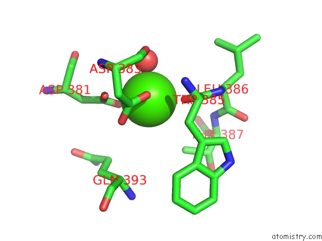
Mono view

Stereo pair view
Mono view
Stereo pair view
A full contact list of Calcium with other atoms in the Ca binding
site number 3 of Crystal Structure of Recombinant GAMMAD364A Fibrinogen Fragment D with the Peptide Ligand Gly-Pro-Arg-Pro-Amide within 5.0Å range:
|
Calcium binding site 4 out of 4 in 3bvh
Go back to
Calcium binding site 4 out
of 4 in the Crystal Structure of Recombinant GAMMAD364A Fibrinogen Fragment D with the Peptide Ligand Gly-Pro-Arg-Pro-Amide
Mono view

Stereo pair view
Mono view
Stereo pair view
A full contact list of Calcium with other atoms in the Ca binding
site number 4 of Crystal Structure of Recombinant GAMMAD364A Fibrinogen Fragment D with the Peptide Ligand Gly-Pro-Arg-Pro-Amide within 5.0Å range:
|
Reference:
S.R.Bowley,
B.K.Merenbloom,
N.Okumura,
L.Betts,
A.Heroux,
O.V.Gorkun,
S.T.Lord.
Polymerization-Defective Fibrinogen Variant GAMMAD364A Binds Knob "A" Peptide Mimic. Biochemistry V. 47 8607 2008.
ISSN: ISSN 0006-2960
PubMed: 18642883
DOI: 10.1021/BI8000769
Page generated: Tue Jul 8 11:14:03 2025
ISSN: ISSN 0006-2960
PubMed: 18642883
DOI: 10.1021/BI8000769
Last articles
Cl in 5RA2Cl in 5RA1
Cl in 5R9Z
Cl in 5RA0
Cl in 5R9Y
Cl in 5R9X
Cl in 5R9W
Cl in 5R9V
Cl in 5R9S
Cl in 5R9U