Calcium »
PDB 3bxl-3cfw »
3c22 »
Calcium in PDB 3c22: Crystal Structure of the Carbohydrate Recognition Domain of Human Langerin
Protein crystallography data
The structure of Crystal Structure of the Carbohydrate Recognition Domain of Human Langerin, PDB code: 3c22
was solved by
M.Thepaut,
with X-Ray Crystallography technique. A brief refinement statistics is given in the table below:
| Resolution Low / High (Å) | 47.72 / 1.50 |
| Space group | P 42 |
| Cell size a, b, c (Å), α, β, γ (°) | 79.555, 79.555, 90.144, 90.00, 90.00, 90.00 |
| R / Rfree (%) | 18.9 / 23.6 |
Other elements in 3c22:
The structure of Crystal Structure of the Carbohydrate Recognition Domain of Human Langerin also contains other interesting chemical elements:
| Magnesium | (Mg) | 6 atoms |
Calcium Binding Sites:
The binding sites of Calcium atom in the Crystal Structure of the Carbohydrate Recognition Domain of Human Langerin
(pdb code 3c22). This binding sites where shown within
5.0 Angstroms radius around Calcium atom.
In total 4 binding sites of Calcium where determined in the Crystal Structure of the Carbohydrate Recognition Domain of Human Langerin, PDB code: 3c22:
Jump to Calcium binding site number: 1; 2; 3; 4;
In total 4 binding sites of Calcium where determined in the Crystal Structure of the Carbohydrate Recognition Domain of Human Langerin, PDB code: 3c22:
Jump to Calcium binding site number: 1; 2; 3; 4;
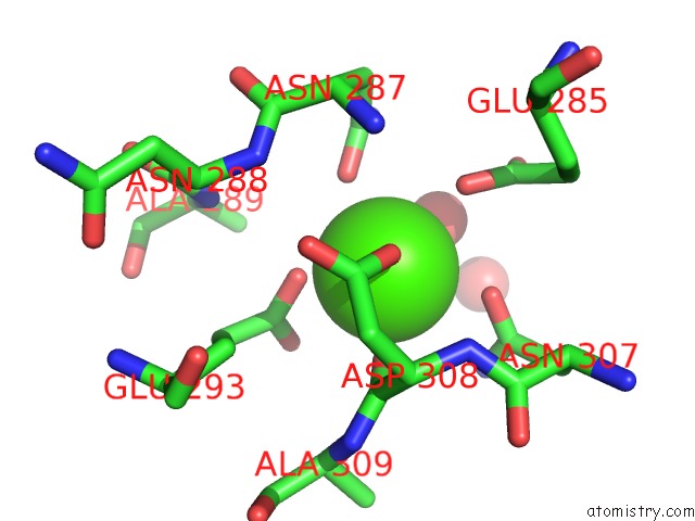
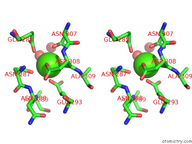
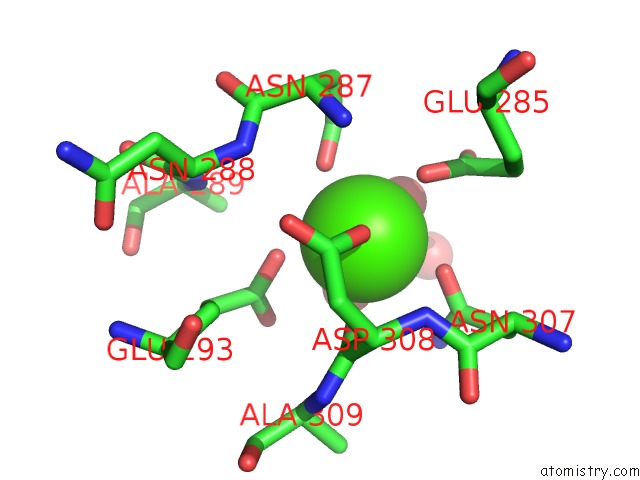
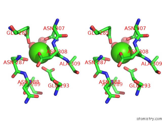
Calcium binding site 1 out of 4 in 3c22
Go back to
Calcium binding site 1 out
of 4 in the Crystal Structure of the Carbohydrate Recognition Domain of Human Langerin
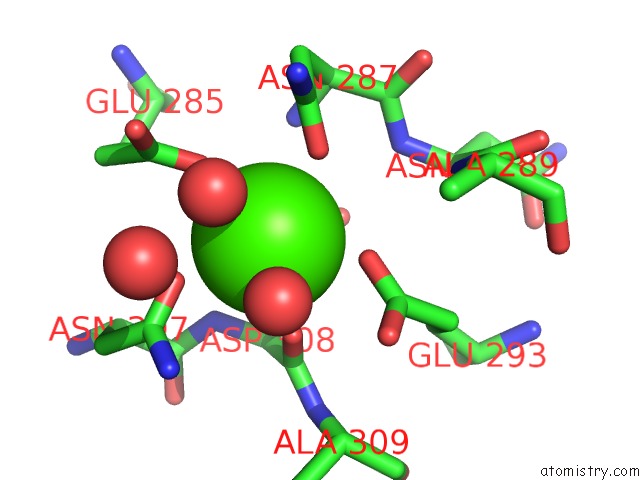
Mono view
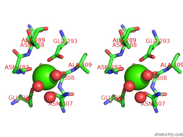
Stereo pair view
Mono view
Stereo pair view
A full contact list of Calcium with other atoms in the Ca binding
site number 1 of Crystal Structure of the Carbohydrate Recognition Domain of Human Langerin within 5.0Å range:
|
Calcium binding site 2 out of 4 in 3c22
Go back to
Calcium binding site 2 out
of 4 in the Crystal Structure of the Carbohydrate Recognition Domain of Human Langerin
Mono view
Stereo pair view
Mono view
Stereo pair view
A full contact list of Calcium with other atoms in the Ca binding
site number 2 of Crystal Structure of the Carbohydrate Recognition Domain of Human Langerin within 5.0Å range:
|
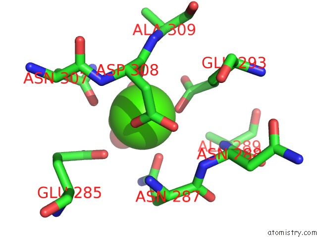
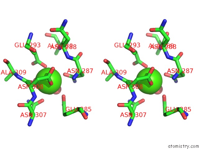
Calcium binding site 3 out of 4 in 3c22
Go back to
Calcium binding site 3 out
of 4 in the Crystal Structure of the Carbohydrate Recognition Domain of Human Langerin
Mono view
Stereo pair view
Mono view
Stereo pair view
A full contact list of Calcium with other atoms in the Ca binding
site number 3 of Crystal Structure of the Carbohydrate Recognition Domain of Human Langerin within 5.0Å range:
|
Calcium binding site 4 out of 4 in 3c22
Go back to
Calcium binding site 4 out
of 4 in the Crystal Structure of the Carbohydrate Recognition Domain of Human Langerin
Mono view
Stereo pair view
Mono view
Stereo pair view
A full contact list of Calcium with other atoms in the Ca binding
site number 4 of Crystal Structure of the Carbohydrate Recognition Domain of Human Langerin within 5.0Å range:
|
Reference:
M.Thepaut,
J.Valladeau,
A.Nurisso,
R.Kahn,
B.Arnou,
C.Vives,
S.Saeland,
C.Ebel,
C.Monnier,
C.Dezutter-Dambuyant,
A.Imberty,
F.Fieschi.
Structural Studies of Langerin and Birbeck Granule: A Macromolecular Organization Model Biochemistry V. 48 2684 2009.
ISSN: ISSN 0006-2960
PubMed: 19175323
DOI: 10.1021/BI802151W
Page generated: Sat Jul 13 08:32:39 2024
ISSN: ISSN 0006-2960
PubMed: 19175323
DOI: 10.1021/BI802151W
Last articles
Zn in 9J0NZn in 9J0O
Zn in 9J0P
Zn in 9FJX
Zn in 9EKB
Zn in 9C0F
Zn in 9CAH
Zn in 9CH0
Zn in 9CH3
Zn in 9CH1