Calcium »
PDB 3bxl-3cfw »
3c6l »
Calcium in PDB 3c6l: Crystal Structure of Mouse Mhc Class II I-Ab/3K Peptide Complexed with Mouse Tcr 2W20
Protein crystallography data
The structure of Crystal Structure of Mouse Mhc Class II I-Ab/3K Peptide Complexed with Mouse Tcr 2W20, PDB code: 3c6l
was solved by
S.Dai,
with X-Ray Crystallography technique. A brief refinement statistics is given in the table below:
| Resolution Low / High (Å) | 49.07 / 3.40 |
| Space group | P 21 21 21 |
| Cell size a, b, c (Å), α, β, γ (°) | 46.540, 113.930, 386.260, 90.00, 90.00, 90.00 |
| R / Rfree (%) | 26.8 / 32.5 |
Calcium Binding Sites:
The binding sites of Calcium atom in the Crystal Structure of Mouse Mhc Class II I-Ab/3K Peptide Complexed with Mouse Tcr 2W20
(pdb code 3c6l). This binding sites where shown within
5.0 Angstroms radius around Calcium atom.
In total 3 binding sites of Calcium where determined in the Crystal Structure of Mouse Mhc Class II I-Ab/3K Peptide Complexed with Mouse Tcr 2W20, PDB code: 3c6l:
Jump to Calcium binding site number: 1; 2; 3;
In total 3 binding sites of Calcium where determined in the Crystal Structure of Mouse Mhc Class II I-Ab/3K Peptide Complexed with Mouse Tcr 2W20, PDB code: 3c6l:
Jump to Calcium binding site number: 1; 2; 3;
Calcium binding site 1 out of 3 in 3c6l
Go back to
Calcium binding site 1 out
of 3 in the Crystal Structure of Mouse Mhc Class II I-Ab/3K Peptide Complexed with Mouse Tcr 2W20
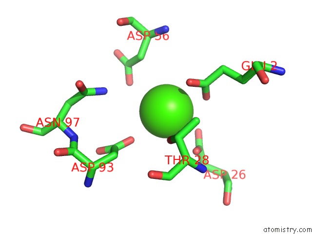
Mono view
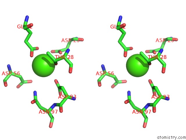
Stereo pair view
Mono view
Stereo pair view
A full contact list of Calcium with other atoms in the Ca binding
site number 1 of Crystal Structure of Mouse Mhc Class II I-Ab/3K Peptide Complexed with Mouse Tcr 2W20 within 5.0Å range:
|
Calcium binding site 2 out of 3 in 3c6l
Go back to
Calcium binding site 2 out
of 3 in the Crystal Structure of Mouse Mhc Class II I-Ab/3K Peptide Complexed with Mouse Tcr 2W20
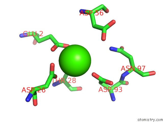
Mono view
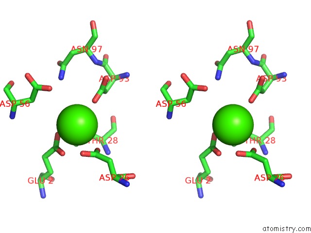
Stereo pair view
Mono view
Stereo pair view
A full contact list of Calcium with other atoms in the Ca binding
site number 2 of Crystal Structure of Mouse Mhc Class II I-Ab/3K Peptide Complexed with Mouse Tcr 2W20 within 5.0Å range:
|
Calcium binding site 3 out of 3 in 3c6l
Go back to
Calcium binding site 3 out
of 3 in the Crystal Structure of Mouse Mhc Class II I-Ab/3K Peptide Complexed with Mouse Tcr 2W20
Mono view
Stereo pair view
Mono view
Stereo pair view
| A full contact list of Calcium with other atoms in the Ca binding site number 3 of Crystal Structure of Mouse Mhc Class II I-Ab/3K Peptide Complexed with Mouse Tcr 2W20 within 5.0Å range: |
Reference:
S.Dai,
E.S.Huseby,
K.Rubtsova,
J.Scott-Browne,
F.Crawford,
W.A.Macdonald,
P.Marrack,
J.W.Kappler.
Crossreactive T Cells Spotlight the Germline Rules For Alphabeta T Cell-Receptor Interactions with Mhc Molecules. Immunity V. 28 324 2008.
ISSN: ISSN 1074-7613
PubMed: 18308592
DOI: 10.1016/J.IMMUNI.2008.01.008
Page generated: Sat Jul 13 08:34:52 2024
ISSN: ISSN 1074-7613
PubMed: 18308592
DOI: 10.1016/J.IMMUNI.2008.01.008
Last articles
Zn in 9J0NZn in 9J0O
Zn in 9J0P
Zn in 9FJX
Zn in 9EKB
Zn in 9C0F
Zn in 9CAH
Zn in 9CH0
Zn in 9CH3
Zn in 9CH1