Calcium »
PDB 3bxl-3cfw »
3c9e »
Calcium in PDB 3c9e: Crystal Structure of the Cathepsin K : Chondroitin Sulfate Complex.
Enzymatic activity of Crystal Structure of the Cathepsin K : Chondroitin Sulfate Complex.
All present enzymatic activity of Crystal Structure of the Cathepsin K : Chondroitin Sulfate Complex.:
3.4.22.38;
3.4.22.38;
Protein crystallography data
The structure of Crystal Structure of the Cathepsin K : Chondroitin Sulfate Complex., PDB code: 3c9e
was solved by
M.Kienetz,
M.M.Cherney,
M.N.G.James,
D.Bromme,
with X-Ray Crystallography technique. A brief refinement statistics is given in the table below:
| Resolution Low / High (Å) | 40.00 / 1.80 |
| Space group | C 2 2 21 |
| Cell size a, b, c (Å), α, β, γ (°) | 42.000, 143.900, 87.200, 90.00, 90.00, 90.00 |
| R / Rfree (%) | 18 / 20.9 |
Calcium Binding Sites:
The binding sites of Calcium atom in the Crystal Structure of the Cathepsin K : Chondroitin Sulfate Complex.
(pdb code 3c9e). This binding sites where shown within
5.0 Angstroms radius around Calcium atom.
In total only one binding site of Calcium was determined in the Crystal Structure of the Cathepsin K : Chondroitin Sulfate Complex., PDB code: 3c9e:
In total only one binding site of Calcium was determined in the Crystal Structure of the Cathepsin K : Chondroitin Sulfate Complex., PDB code: 3c9e:
Calcium binding site 1 out of 1 in 3c9e
Go back to
Calcium binding site 1 out
of 1 in the Crystal Structure of the Cathepsin K : Chondroitin Sulfate Complex.
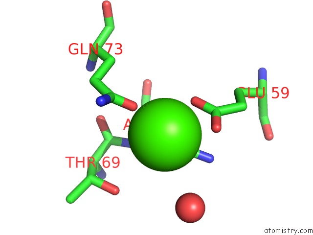
Mono view
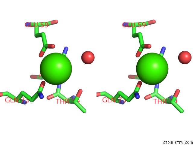
Stereo pair view
Mono view
Stereo pair view
A full contact list of Calcium with other atoms in the Ca binding
site number 1 of Crystal Structure of the Cathepsin K : Chondroitin Sulfate Complex. within 5.0Å range:
|
Reference:
Z.Li,
M.Kienetz,
M.M.Cherney,
M.N.James,
D.Bromme.
The Crystal and Molecular Structures of A Cathepsin K:Chondroitin Sulfate Complex. J.Mol.Biol. V. 383 78 2008.
ISSN: ISSN 0022-2836
PubMed: 18692071
DOI: 10.1016/J.JMB.2008.07.038
Page generated: Sat Jul 13 08:36:42 2024
ISSN: ISSN 0022-2836
PubMed: 18692071
DOI: 10.1016/J.JMB.2008.07.038
Last articles
Zn in 9J0NZn in 9J0O
Zn in 9J0P
Zn in 9FJX
Zn in 9EKB
Zn in 9C0F
Zn in 9CAH
Zn in 9CH0
Zn in 9CH3
Zn in 9CH1