Calcium »
PDB 3cga-3ctz »
3cga »
Calcium in PDB 3cga: Crystal Structure of Metastasis-Associated Protein S100A4 in the Active, Calcium-Bound Form
Protein crystallography data
The structure of Crystal Structure of Metastasis-Associated Protein S100A4 in the Active, Calcium-Bound Form, PDB code: 3cga
was solved by
P.Pathuri,
H.Luecke,
with X-Ray Crystallography technique. A brief refinement statistics is given in the table below:
| Resolution Low / High (Å) | 50.00 / 2.03 |
| Space group | P 65 |
| Cell size a, b, c (Å), α, β, γ (°) | 47.107, 47.107, 176.200, 90.00, 90.00, 120.00 |
| R / Rfree (%) | 25.5 / 33.1 |
Calcium Binding Sites:
The binding sites of Calcium atom in the Crystal Structure of Metastasis-Associated Protein S100A4 in the Active, Calcium-Bound Form
(pdb code 3cga). This binding sites where shown within
5.0 Angstroms radius around Calcium atom.
In total 4 binding sites of Calcium where determined in the Crystal Structure of Metastasis-Associated Protein S100A4 in the Active, Calcium-Bound Form, PDB code: 3cga:
Jump to Calcium binding site number: 1; 2; 3; 4;
In total 4 binding sites of Calcium where determined in the Crystal Structure of Metastasis-Associated Protein S100A4 in the Active, Calcium-Bound Form, PDB code: 3cga:
Jump to Calcium binding site number: 1; 2; 3; 4;
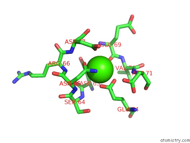
Calcium binding site 1 out of 4 in 3cga
Go back to
Calcium binding site 1 out
of 4 in the Crystal Structure of Metastasis-Associated Protein S100A4 in the Active, Calcium-Bound Form
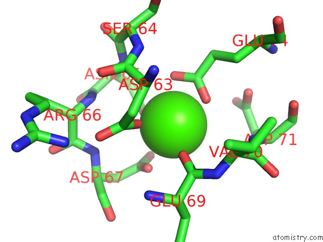
Mono view

Stereo pair view
Mono view
Stereo pair view
A full contact list of Calcium with other atoms in the Ca binding
site number 1 of Crystal Structure of Metastasis-Associated Protein S100A4 in the Active, Calcium-Bound Form within 5.0Å range:
|
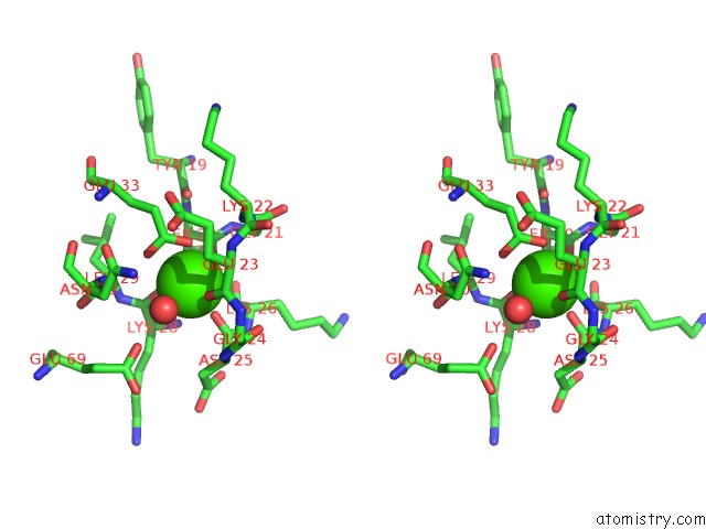
Calcium binding site 2 out of 4 in 3cga
Go back to
Calcium binding site 2 out
of 4 in the Crystal Structure of Metastasis-Associated Protein S100A4 in the Active, Calcium-Bound Form

Mono view
Stereo pair view
Mono view
Stereo pair view
A full contact list of Calcium with other atoms in the Ca binding
site number 2 of Crystal Structure of Metastasis-Associated Protein S100A4 in the Active, Calcium-Bound Form within 5.0Å range:
|
Calcium binding site 3 out of 4 in 3cga
Go back to
Calcium binding site 3 out
of 4 in the Crystal Structure of Metastasis-Associated Protein S100A4 in the Active, Calcium-Bound Form
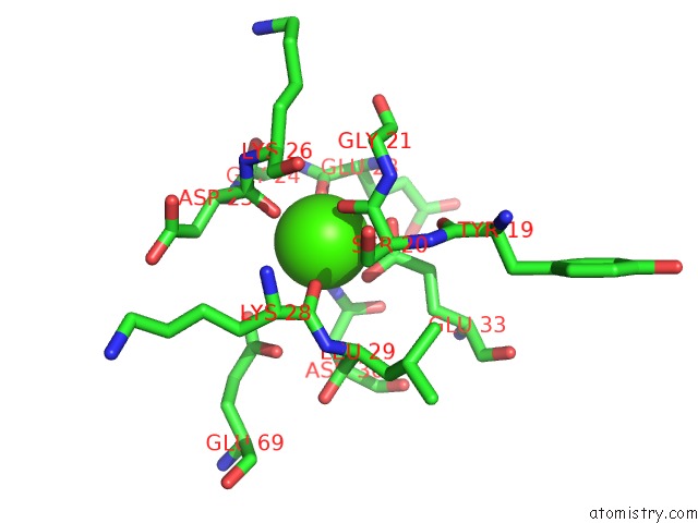
Mono view
Stereo pair view
Mono view
Stereo pair view
A full contact list of Calcium with other atoms in the Ca binding
site number 3 of Crystal Structure of Metastasis-Associated Protein S100A4 in the Active, Calcium-Bound Form within 5.0Å range:
|
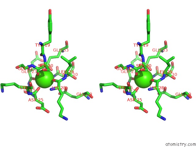
Calcium binding site 4 out of 4 in 3cga
Go back to
Calcium binding site 4 out
of 4 in the Crystal Structure of Metastasis-Associated Protein S100A4 in the Active, Calcium-Bound Form
Mono view

Stereo pair view
Mono view
Stereo pair view
A full contact list of Calcium with other atoms in the Ca binding
site number 4 of Crystal Structure of Metastasis-Associated Protein S100A4 in the Active, Calcium-Bound Form within 5.0Å range:
|
Reference:
P.Pathuri,
L.Vogeley,
H.Luecke.
Crystal Structure of Metastasis-Associated Protein S100A4 in the Active Calcium-Bound Form J.Mol.Biol. V. 383 62 2008.
ISSN: ISSN 0022-2836
PubMed: 18783790
DOI: 10.1016/J.JMB.2008.04.076
Page generated: Sat Jul 13 08:38:54 2024
ISSN: ISSN 0022-2836
PubMed: 18783790
DOI: 10.1016/J.JMB.2008.04.076
Last articles
Zn in 9J0NZn in 9J0O
Zn in 9J0P
Zn in 9FJX
Zn in 9EKB
Zn in 9C0F
Zn in 9CAH
Zn in 9CH0
Zn in 9CH3
Zn in 9CH1