Calcium »
PDB 3cga-3ctz »
3cjb »
Calcium in PDB 3cjb: Actin Dimer Cross-Linked By V. Cholerae Martx Toxin and Complexed with Gelsolin-Segment 1
Protein crystallography data
The structure of Actin Dimer Cross-Linked By V. Cholerae Martx Toxin and Complexed with Gelsolin-Segment 1, PDB code: 3cjb
was solved by
M.R.Sawaya,
D.S.Kudryashov,
I.Pashkov,
E.Reisler,
T.O.Yeates,
with X-Ray Crystallography technique. A brief refinement statistics is given in the table below:
| Resolution Low / High (Å) | 90.91 / 3.21 |
| Space group | P 21 21 21 |
| Cell size a, b, c (Å), α, β, γ (°) | 56.696, 69.540, 182.106, 90.00, 90.00, 90.00 |
| R / Rfree (%) | 24.5 / 26.9 |
Calcium Binding Sites:
The binding sites of Calcium atom in the Actin Dimer Cross-Linked By V. Cholerae Martx Toxin and Complexed with Gelsolin-Segment 1
(pdb code 3cjb). This binding sites where shown within
5.0 Angstroms radius around Calcium atom.
In total 2 binding sites of Calcium where determined in the Actin Dimer Cross-Linked By V. Cholerae Martx Toxin and Complexed with Gelsolin-Segment 1, PDB code: 3cjb:
Jump to Calcium binding site number: 1; 2;
In total 2 binding sites of Calcium where determined in the Actin Dimer Cross-Linked By V. Cholerae Martx Toxin and Complexed with Gelsolin-Segment 1, PDB code: 3cjb:
Jump to Calcium binding site number: 1; 2;
Calcium binding site 1 out of 2 in 3cjb
Go back to
Calcium binding site 1 out
of 2 in the Actin Dimer Cross-Linked By V. Cholerae Martx Toxin and Complexed with Gelsolin-Segment 1
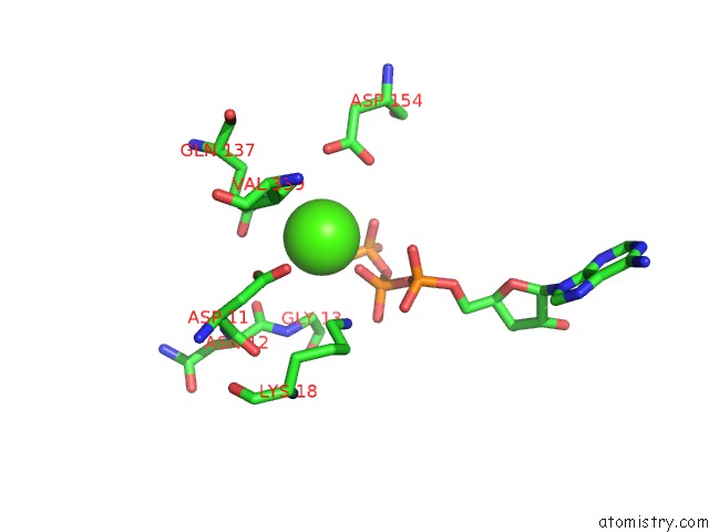
Mono view
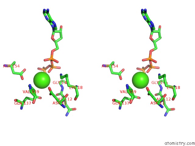
Stereo pair view
Mono view
Stereo pair view
A full contact list of Calcium with other atoms in the Ca binding
site number 1 of Actin Dimer Cross-Linked By V. Cholerae Martx Toxin and Complexed with Gelsolin-Segment 1 within 5.0Å range:
|
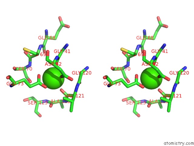
Calcium binding site 2 out of 2 in 3cjb
Go back to
Calcium binding site 2 out
of 2 in the Actin Dimer Cross-Linked By V. Cholerae Martx Toxin and Complexed with Gelsolin-Segment 1
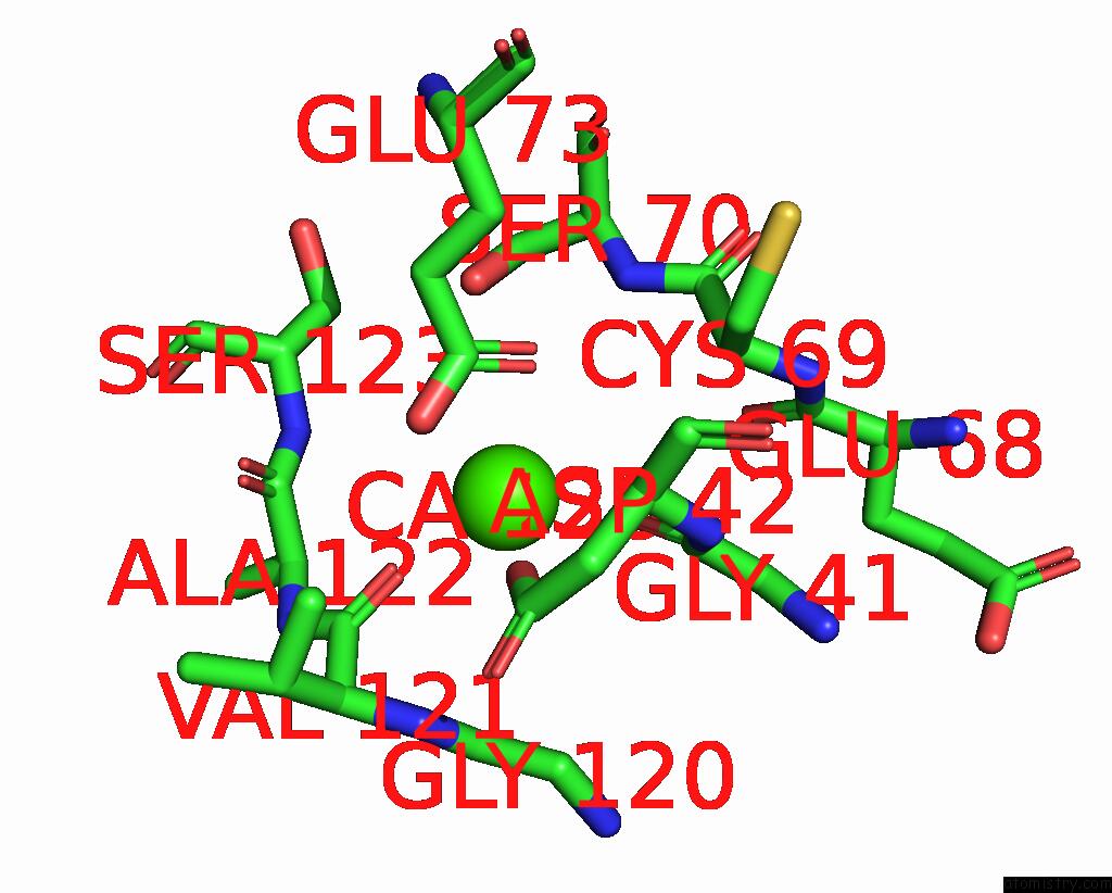
Mono view
Stereo pair view
Mono view
Stereo pair view
A full contact list of Calcium with other atoms in the Ca binding
site number 2 of Actin Dimer Cross-Linked By V. Cholerae Martx Toxin and Complexed with Gelsolin-Segment 1 within 5.0Å range:
|
Reference:
D.S.Kudryashov,
Z.A.Durer,
A.J.Ytterberg,
M.R.Sawaya,
I.Pashkov,
K.Prochazkova,
T.O.Yeates,
R.R.Loo,
J.A.Loo,
K.J.Satchell,
E.Reisler.
Connecting Actin Monomers By Iso-Peptide Bond Is A Toxicity Mechanism of the Vibrio Cholerae Martx Toxin. Proc.Natl.Acad.Sci.Usa V. 105 18537 2008.
ISSN: ISSN 0027-8424
PubMed: 19015515
DOI: 10.1073/PNAS.0808082105
Page generated: Sat Jul 13 08:39:55 2024
ISSN: ISSN 0027-8424
PubMed: 19015515
DOI: 10.1073/PNAS.0808082105
Last articles
Zn in 9MJ5Zn in 9HNW
Zn in 9G0L
Zn in 9FNE
Zn in 9DZN
Zn in 9E0I
Zn in 9D32
Zn in 9DAK
Zn in 8ZXC
Zn in 8ZUF