Calcium »
PDB 3cga-3ctz »
3csh »
Calcium in PDB 3csh: Crystal Structure of Glutathione Transferase Pi in Complex with the Chlorambucil-Glutathione Conjugate
Enzymatic activity of Crystal Structure of Glutathione Transferase Pi in Complex with the Chlorambucil-Glutathione Conjugate
All present enzymatic activity of Crystal Structure of Glutathione Transferase Pi in Complex with the Chlorambucil-Glutathione Conjugate:
2.5.1.18;
2.5.1.18;
Protein crystallography data
The structure of Crystal Structure of Glutathione Transferase Pi in Complex with the Chlorambucil-Glutathione Conjugate, PDB code: 3csh
was solved by
L.J.Parker,
with X-Ray Crystallography technique. A brief refinement statistics is given in the table below:
| Resolution Low / High (Å) | 18.69 / 1.55 |
| Space group | C 1 2 1 |
| Cell size a, b, c (Å), α, β, γ (°) | 77.346, 89.358, 68.946, 90.00, 98.03, 90.00 |
| R / Rfree (%) | 17 / 20 |
Other elements in 3csh:
The structure of Crystal Structure of Glutathione Transferase Pi in Complex with the Chlorambucil-Glutathione Conjugate also contains other interesting chemical elements:
| Chlorine | (Cl) | 4 atoms |
Calcium Binding Sites:
The binding sites of Calcium atom in the Crystal Structure of Glutathione Transferase Pi in Complex with the Chlorambucil-Glutathione Conjugate
(pdb code 3csh). This binding sites where shown within
5.0 Angstroms radius around Calcium atom.
In total only one binding site of Calcium was determined in the Crystal Structure of Glutathione Transferase Pi in Complex with the Chlorambucil-Glutathione Conjugate, PDB code: 3csh:
In total only one binding site of Calcium was determined in the Crystal Structure of Glutathione Transferase Pi in Complex with the Chlorambucil-Glutathione Conjugate, PDB code: 3csh:
Calcium binding site 1 out of 1 in 3csh
Go back to
Calcium binding site 1 out
of 1 in the Crystal Structure of Glutathione Transferase Pi in Complex with the Chlorambucil-Glutathione Conjugate
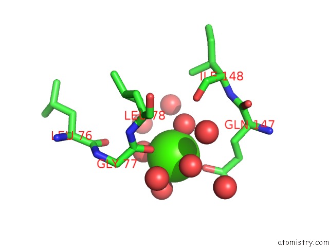
Mono view
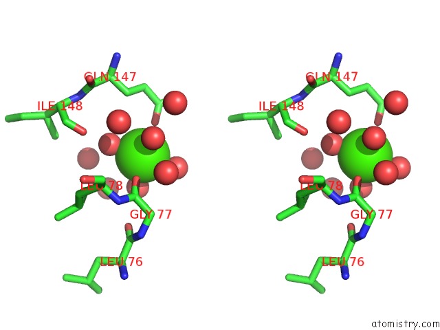
Stereo pair view
Mono view
Stereo pair view
A full contact list of Calcium with other atoms in the Ca binding
site number 1 of Crystal Structure of Glutathione Transferase Pi in Complex with the Chlorambucil-Glutathione Conjugate within 5.0Å range:
|
Reference:
L.J.Parker,
S.Ciccone,
L.C.Italiano,
A.Primavera,
A.J.Oakley,
C.J.Morton,
N.C.Hancock,
M.L.Bello,
M.W.Parker.
The Anti-Cancer Drug Chlorambucil As A Substrate For the Human Polymorphic Enzyme Glutathione Transferase P1-1: Kinetic Properties and Crystallographic Characterisation of Allelic Variants. J.Mol.Biol. V. 380 131 2008.
ISSN: ISSN 0022-2836
PubMed: 18511072
DOI: 10.1016/J.JMB.2008.04.066
Page generated: Sat Jul 13 08:45:51 2024
ISSN: ISSN 0022-2836
PubMed: 18511072
DOI: 10.1016/J.JMB.2008.04.066
Last articles
Zn in 9J0NZn in 9J0O
Zn in 9J0P
Zn in 9FJX
Zn in 9EKB
Zn in 9C0F
Zn in 9CAH
Zn in 9CH0
Zn in 9CH3
Zn in 9CH1