Calcium »
PDB 3cu2-3dc0 »
3cv1 »
Calcium in PDB 3cv1: Atomic Resolution Structures of Escherichia Coli and Bacillis Anthracis Malate Synthase A: Comparison with Isoform G and Implications For Structure Based Drug Design
Enzymatic activity of Atomic Resolution Structures of Escherichia Coli and Bacillis Anthracis Malate Synthase A: Comparison with Isoform G and Implications For Structure Based Drug Design
All present enzymatic activity of Atomic Resolution Structures of Escherichia Coli and Bacillis Anthracis Malate Synthase A: Comparison with Isoform G and Implications For Structure Based Drug Design:
2.3.3.9;
2.3.3.9;
Protein crystallography data
The structure of Atomic Resolution Structures of Escherichia Coli and Bacillis Anthracis Malate Synthase A: Comparison with Isoform G and Implications For Structure Based Drug Design, PDB code: 3cv1
was solved by
J.R.Lohman,
with X-Ray Crystallography technique. A brief refinement statistics is given in the table below:
| Resolution Low / High (Å) | 30.00 / 1.68 |
| Space group | P 1 21 1 |
| Cell size a, b, c (Å), α, β, γ (°) | 52.031, 74.124, 71.917, 90.00, 97.82, 90.00 |
| R / Rfree (%) | 17.6 / 20.9 |
Calcium Binding Sites:
The binding sites of Calcium atom in the Atomic Resolution Structures of Escherichia Coli and Bacillis Anthracis Malate Synthase A: Comparison with Isoform G and Implications For Structure Based Drug Design
(pdb code 3cv1). This binding sites where shown within
5.0 Angstroms radius around Calcium atom.
In total 2 binding sites of Calcium where determined in the Atomic Resolution Structures of Escherichia Coli and Bacillis Anthracis Malate Synthase A: Comparison with Isoform G and Implications For Structure Based Drug Design, PDB code: 3cv1:
Jump to Calcium binding site number: 1; 2;
In total 2 binding sites of Calcium where determined in the Atomic Resolution Structures of Escherichia Coli and Bacillis Anthracis Malate Synthase A: Comparison with Isoform G and Implications For Structure Based Drug Design, PDB code: 3cv1:
Jump to Calcium binding site number: 1; 2;
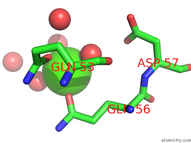
Calcium binding site 1 out of 2 in 3cv1
Go back to
Calcium binding site 1 out
of 2 in the Atomic Resolution Structures of Escherichia Coli and Bacillis Anthracis Malate Synthase A: Comparison with Isoform G and Implications For Structure Based Drug Design
Mono view
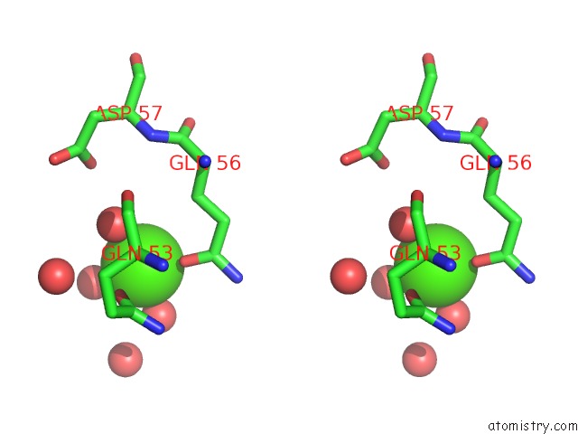
Stereo pair view
Mono view
Stereo pair view
A full contact list of Calcium with other atoms in the Ca binding
site number 1 of Atomic Resolution Structures of Escherichia Coli and Bacillis Anthracis Malate Synthase A: Comparison with Isoform G and Implications For Structure Based Drug Design within 5.0Å range:
|
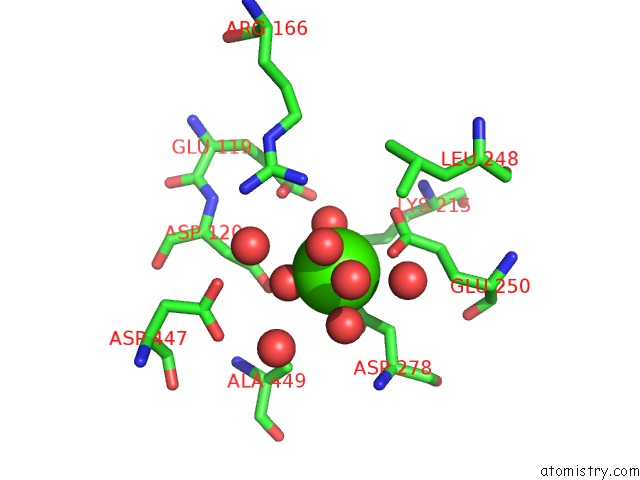
Calcium binding site 2 out of 2 in 3cv1
Go back to
Calcium binding site 2 out
of 2 in the Atomic Resolution Structures of Escherichia Coli and Bacillis Anthracis Malate Synthase A: Comparison with Isoform G and Implications For Structure Based Drug Design
Mono view
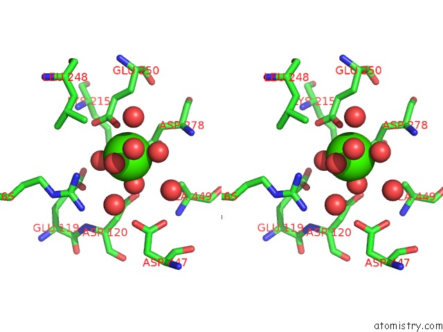
Stereo pair view
Mono view
Stereo pair view
A full contact list of Calcium with other atoms in the Ca binding
site number 2 of Atomic Resolution Structures of Escherichia Coli and Bacillis Anthracis Malate Synthase A: Comparison with Isoform G and Implications For Structure Based Drug Design within 5.0Å range:
|
Reference:
J.R.Lohman,
A.C.Olson,
S.J.Remington.
Atomic Resolution Structures of Escherichia Coli and Bacillus Anthracis Malate Synthase A: Comparison with Isoform G and Implications For Structure-Based Drug Discovery Protein Sci. V. 17 1935 2008.
ISSN: ISSN 0961-8368
PubMed: 18714089
DOI: 10.1110/PS.036269.108
Page generated: Tue Jul 8 11:27:50 2025
ISSN: ISSN 0961-8368
PubMed: 18714089
DOI: 10.1110/PS.036269.108
Last articles
Cl in 8D58Cl in 8D5R
Cl in 8D5G
Cl in 8D5H
Cl in 8D4Z
Cl in 8D5B
Cl in 8D4I
Cl in 8D59
Cl in 8D4P
Cl in 8D3S