Atomistry » Calcium » PDB 3cu2-3dc0 » 3d10
Atomistry »
  Calcium »
    PDB 3cu2-3dc0 »
      3d10 »

Calcium in PDB 3d10: Crystal Structure of S100B in the Calcium and Zinc Loaded State at pH 10.0

Protein crystallography data

The structure of Crystal Structure of S100B in the Calcium and Zinc Loaded State at pH 10.0, PDB code: 3d10 was solved by T.Ostendorp, J.Diez, C.W.Heizmann, G.Fritz, with X-Ray Crystallography technique. A brief refinement statistics is given in the table below:

Resolution Low / High (Å) 10.00 / 1.65
Space group P 1 21 1
Cell size a, b, c (Å), α, β, γ (°) 34.750, 58.180, 47.850, 90.00, 111.08, 90.00
R / Rfree (%) 20.9 / 27.5

Other elements in 3d10:

The structure of Crystal Structure of S100B in the Calcium and Zinc Loaded State at pH 10.0 also contains other interesting chemical elements:

Zinc (Zn) 2 atoms

Calcium Binding Sites:

The binding sites of Calcium atom in the Crystal Structure of S100B in the Calcium and Zinc Loaded State at pH 10.0 (pdb code 3d10). This binding sites where shown within 5.0 Angstroms radius around Calcium atom.
In total 4 binding sites of Calcium where determined in the Crystal Structure of S100B in the Calcium and Zinc Loaded State at pH 10.0, PDB code: 3d10:
Jump to Calcium binding site number: 1; 2; 3; 4;

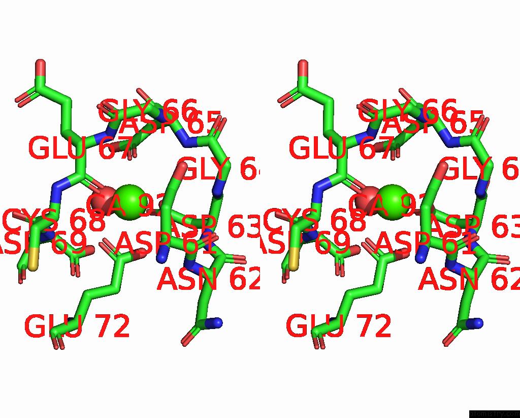
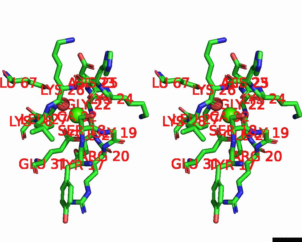
Calcium binding site 1 out of 4 in 3d10

Go back to Calcium Binding Sites List in 3d10
Calcium binding site 1 out of 4 in the Crystal Structure of S100B in the Calcium and Zinc Loaded State at pH 10.0


Mono view


Stereo pair view

A full contact list of Calcium with other atoms in the Ca binding site number 1 of Crystal Structure of S100B in the Calcium and Zinc Loaded State at pH 10.0 within 5.0Å range:
probe atom residue distance (Å) B Occ
A:Ca92

b:30.2
occ:1.00
O A:GLU67 2.2 10.2 1.0
OD2 A:ASP63 2.3 15.1 1.0
OD1 A:ASP61 2.3 18.4 1.0
O A:HOH98 2.3 32.7 1.0
OD1 A:ASP65 2.4 12.2 1.0
OE1 A:GLU72 2.4 11.9 1.0
OE2 A:GLU72 2.7 10.2 1.0
CD A:GLU72 2.9 13.8 1.0
CG A:ASP63 3.3 19.7 1.0
CG A:ASP61 3.3 20.1 1.0
C A:GLU67 3.4 12.3 1.0
CG A:ASP65 3.4 18.6 1.0
OD1 A:ASP63 3.6 23.5 1.0
OD2 A:ASP65 3.9 17.6 1.0
N A:GLU67 4.0 14.3 1.0
OD2 A:ASP61 4.1 16.3 1.0
CB A:ASP61 4.2 20.1 1.0
N A:ASP65 4.2 19.9 1.0
CA A:GLU67 4.2 12.4 1.0
N A:CYS68 4.3 11.2 1.0
CA A:ASP61 4.3 19.5 1.0
CA A:CYS68 4.3 11.9 1.0
N A:ASP63 4.4 19.3 1.0
N A:ASP69 4.4 12.4 1.0
CG A:GLU72 4.4 12.4 1.0
N A:GLY64 4.5 20.5 1.0
CB A:ASP63 4.5 18.3 1.0
CB A:ASP65 4.6 19.3 1.0
N A:ASN62 4.6 18.9 1.0
C A:ASP61 4.7 19.8 1.0
CA A:ASP63 4.7 19.2 1.0
C A:CYS68 4.8 11.9 1.0
C A:ASP63 4.8 19.4 1.0
CA A:ASP65 4.8 19.3 1.0
CB A:GLU67 4.8 13.5 1.0
N A:GLY66 4.8 17.5 1.0
CG A:ASP69 4.9 13.5 1.0
OD2 A:ASP69 4.9 17.2 1.0
C A:ASP65 4.9 18.4 1.0

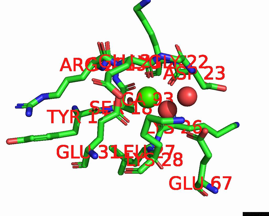
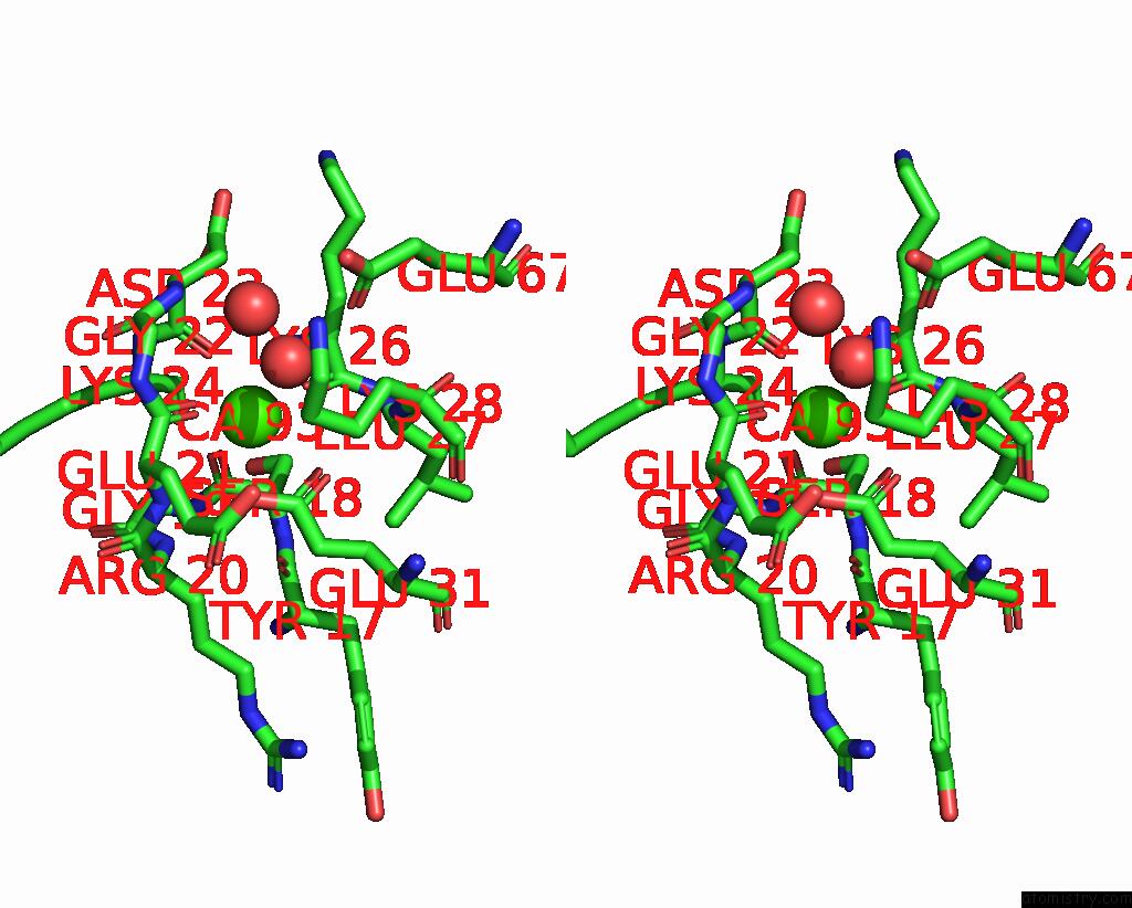
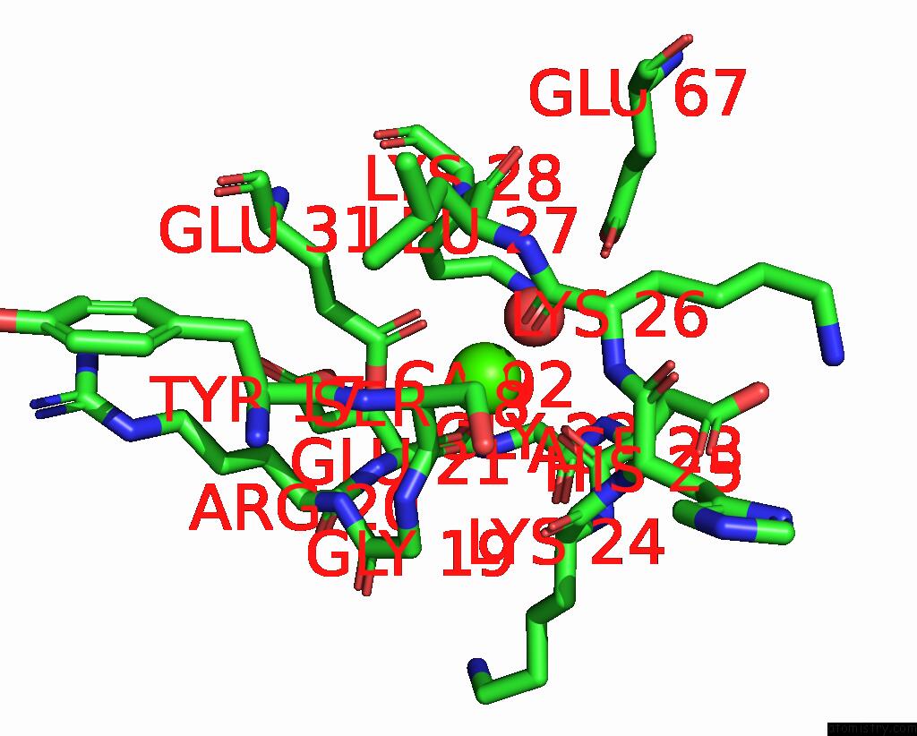
Calcium binding site 2 out of 4 in 3d10

Go back to Calcium Binding Sites List in 3d10
Calcium binding site 2 out of 4 in the Crystal Structure of S100B in the Calcium and Zinc Loaded State at pH 10.0


Mono view


Stereo pair view

A full contact list of Calcium with other atoms in the Ca binding site number 2 of Crystal Structure of S100B in the Calcium and Zinc Loaded State at pH 10.0 within 5.0Å range:
probe atom residue distance (Å) B Occ
A:Ca93

b:33.4
occ:1.00
O A:GLU21 2.3 13.5 1.0
O A:SER18 2.3 13.8 1.0
O A:HOH123 2.4 33.2 1.0
O A:LYS26 2.5 15.4 1.0
O A:ASP23 2.7 20.9 1.0
OE2 A:GLU31 2.7 17.2 1.0
OE1 A:GLU31 2.8 19.9 1.0
CD A:GLU31 3.1 13.4 1.0
C A:SER18 3.3 16.8 1.0
C A:GLU21 3.5 17.0 1.0
C A:LYS26 3.7 15.3 1.0
C A:ASP23 3.7 21.1 1.0
CA A:SER18 3.8 16.7 1.0
N A:GLU21 4.0 18.3 1.0
N A:ASP23 4.3 20.6 1.0
CA A:GLU21 4.3 18.2 1.0
N A:LYS28 4.4 14.9 1.0
C A:GLY22 4.4 19.3 1.0
N A:LYS26 4.4 17.6 1.0
CA A:LYS24 4.5 20.5 1.0
N A:GLY19 4.5 17.2 1.0
CB A:SER18 4.5 18.8 1.0
N A:GLY22 4.5 16.5 1.0
CG A:GLU31 4.5 15.2 1.0
OE1 A:GLU67 4.5 22.7 1.0
N A:LYS24 4.6 20.9 1.0
CA A:ASP23 4.6 21.1 1.0
N A:ARG20 4.6 19.4 1.0
N A:LEU27 4.6 14.4 1.0
O A:TYR17 4.6 15.1 1.0
CA A:LEU27 4.6 15.8 1.0
CA A:LYS26 4.6 15.9 1.0
CA A:GLY22 4.7 18.3 1.0
C A:LYS24 4.7 20.8 1.0
O A:HOH152 4.7 42.2 1.0
O A:GLY22 4.9 19.3 1.0
CA A:GLY19 4.9 18.5 1.0
CG A:LYS28 4.9 17.7 1.0
C A:LEU27 4.9 14.8 1.0
CB A:ASP23 5.0 21.1 1.0
C A:GLY19 5.0 19.2 1.0
CD A:LYS28 5.0 14.6 1.0

Calcium binding site 3 out of 4 in 3d10

Go back to Calcium Binding Sites List in 3d10
Calcium binding site 3 out of 4 in the Crystal Structure of S100B in the Calcium and Zinc Loaded State at pH 10.0


Mono view


Stereo pair view

A full contact list of Calcium with other atoms in the Ca binding site number 3 of Crystal Structure of S100B in the Calcium and Zinc Loaded State at pH 10.0 within 5.0Å range:
probe atom residue distance (Å) B Occ
B:Ca92

b:31.7
occ:1.00
OE1 B:GLU31 2.2 14.9 1.0
O B:GLU21 2.3 15.2 1.0
O B:SER18 2.3 15.1 1.0
O B:ASP23 2.4 13.4 1.0
O B:HOH132 2.4 39.9 1.0
O B:LYS26 2.5 15.8 1.0
OE2 B:GLU31 2.8 17.2 1.0
CD B:GLU31 2.9 15.2 1.0
C B:SER18 3.4 12.1 1.0
C B:GLU21 3.5 17.9 1.0
C B:ASP23 3.6 16.9 1.0
C B:LYS26 3.7 15.9 1.0
CA B:SER18 3.9 13.4 1.0
N B:GLU21 4.1 15.9 1.0
N B:ASP23 4.2 17.9 1.0
C B:GLY22 4.2 18.6 1.0
N B:LYS28 4.3 16.1 1.0
CA B:GLU21 4.4 18.9 1.0
CG B:GLU31 4.4 14.1 1.0
CA B:ASP23 4.4 16.9 1.0
N B:LYS26 4.4 17.3 1.0
CA B:LEU27 4.5 15.7 1.0
N B:GLY22 4.5 17.7 1.0
N B:LYS24 4.5 17.6 1.0
CA B:LYS24 4.5 17.2 1.0
N B:GLY19 4.5 12.5 1.0
O B:GLY22 4.5 20.2 1.0
OE1 B:GLU67 4.5 18.5 1.0
N B:LEU27 4.5 15.4 1.0
CA B:GLY22 4.6 18.8 1.0
CB B:SER18 4.6 13.2 1.0
CG B:LYS28 4.6 17.9 1.0
O B:TYR17 4.7 12.6 1.0
CA B:LYS26 4.7 16.6 1.0
N B:ARG20 4.7 16.1 1.0
C B:LEU27 4.8 16.4 1.0
C B:LYS24 4.8 17.4 1.0
CB B:ASP23 4.8 16.9 1.0
NZ B:LYS28 4.8 18.7 1.0
CA B:GLY19 4.9 14.5 1.0
CB B:GLU21 4.9 19.7 1.0
N B:HIS25 4.9 17.4 1.0

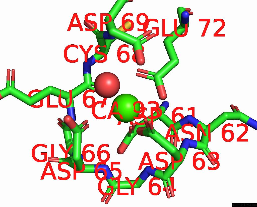
Calcium binding site 4 out of 4 in 3d10

Go back to Calcium Binding Sites List in 3d10
Calcium binding site 4 out of 4 in the Crystal Structure of S100B in the Calcium and Zinc Loaded State at pH 10.0


Mono view


Stereo pair view

A full contact list of Calcium with other atoms in the Ca binding site number 4 of Crystal Structure of S100B in the Calcium and Zinc Loaded State at pH 10.0 within 5.0Å range:
probe atom residue distance (Å) B Occ
B:Ca93

b:35.7
occ:1.00
OD1 B:ASP61 1.8 12.9 1.0
OD2 B:ASP63 2.2 11.0 1.0
O B:GLU67 2.3 9.1 1.0
OE1 B:GLU72 2.4 8.0 1.0
OE2 B:GLU72 2.4 12.1 1.0
OD1 B:ASP65 2.7 16.6 1.0
O B:HOH152 2.7 38.5 1.0
CD B:GLU72 2.8 7.7 1.0
CG B:ASP61 3.1 13.9 1.0
CG B:ASP63 3.3 17.6 1.0
C B:GLU67 3.5 11.2 1.0
CG B:ASP65 3.6 18.2 1.0
OD1 B:ASP63 3.8 21.9 1.0
OD2 B:ASP61 3.9 17.7 1.0
CB B:ASP61 4.0 15.5 1.0
N B:ASP65 4.0 17.3 1.0
CA B:ASP61 4.0 15.0 1.0
N B:GLY64 4.1 17.1 1.0
N B:GLU67 4.2 14.1 1.0
CG B:GLU72 4.3 7.0 1.0
OD2 B:ASP65 4.3 21.0 1.0
N B:ASP63 4.3 16.3 1.0
OD2 B:ASP69 4.3 15.9 1.0
C B:ASP61 4.4 15.9 1.0
CA B:GLU67 4.4 12.7 1.0
N B:CYS68 4.4 10.5 1.0
CA B:CYS68 4.4 10.2 1.0
CB B:ASP65 4.5 17.6 1.0
CB B:ASP63 4.6 16.2 1.0
N B:ASP69 4.6 11.1 1.0
N B:ASN62 4.6 15.1 1.0
CA B:ASP65 4.7 17.2 1.0
CA B:ASP63 4.7 16.3 1.0
CG B:ASP69 4.7 12.2 1.0
N B:GLY66 4.8 15.5 1.0
O B:ASP61 4.8 16.9 1.0
C B:ASP63 4.9 16.8 1.0
CA B:GLY64 5.0 17.8 1.0
C B:GLY64 5.0 18.1 1.0

Reference:

T.Ostendorp, J.Diez, C.W.Heizmann, G.Fritz. The Crystal Structures of Human S100B in the Zinc- and Calcium-Loaded State at Three pH Values Reveal Zinc Ligand Swapping. Biochim.Biophys.Acta V.1813 1083 2011.
ISSN: ISSN 0006-3002
PubMed: 20950652
DOI: 10.1016/J.BBAMCR.2010.10.006
Page generated: Sat Dec 12 04:08:13 2020

Last articles

Zn in 8WB0
Zn in 8WAX
Zn in 8WAU
Zn in 8WAZ
Zn in 8WAY
Zn in 8WAV
Zn in 8WAW
Zn in 8WAT
Zn in 8W7M
Zn in 8WD3
© Copyright 2008-2020 by atomistry.com
Home   |    Site Map   |    Copyright   |    Contact us   |    Privacy