Calcium »
PDB 3dcq-3dsw »
3dhq »
Calcium in PDB 3dhq: Crystal Structure of Staphylococcal Nuclease Variant Delta+Phs A90R at Cryogenic Temperature
Enzymatic activity of Crystal Structure of Staphylococcal Nuclease Variant Delta+Phs A90R at Cryogenic Temperature
All present enzymatic activity of Crystal Structure of Staphylococcal Nuclease Variant Delta+Phs A90R at Cryogenic Temperature:
3.1.31.1;
3.1.31.1;
Protein crystallography data
The structure of Crystal Structure of Staphylococcal Nuclease Variant Delta+Phs A90R at Cryogenic Temperature, PDB code: 3dhq
was solved by
M.J.Harms,
J.L.Schlessman,
E.B.Garcia-Moreno,
with X-Ray Crystallography technique. A brief refinement statistics is given in the table below:
| Resolution Low / High (Å) | 30.30 / 2.15 |
| Space group | P 1 21 1 |
| Cell size a, b, c (Å), α, β, γ (°) | 30.931, 60.603, 38.421, 90.00, 93.93, 90.00 |
| R / Rfree (%) | 20.8 / 27.6 |
Calcium Binding Sites:
The binding sites of Calcium atom in the Crystal Structure of Staphylococcal Nuclease Variant Delta+Phs A90R at Cryogenic Temperature
(pdb code 3dhq). This binding sites where shown within
5.0 Angstroms radius around Calcium atom.
In total only one binding site of Calcium was determined in the Crystal Structure of Staphylococcal Nuclease Variant Delta+Phs A90R at Cryogenic Temperature, PDB code: 3dhq:
In total only one binding site of Calcium was determined in the Crystal Structure of Staphylococcal Nuclease Variant Delta+Phs A90R at Cryogenic Temperature, PDB code: 3dhq:
Calcium binding site 1 out of 1 in 3dhq
Go back to
Calcium binding site 1 out
of 1 in the Crystal Structure of Staphylococcal Nuclease Variant Delta+Phs A90R at Cryogenic Temperature
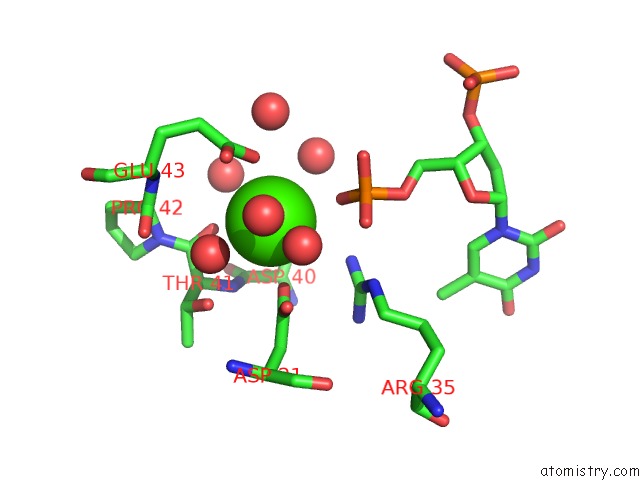
Mono view
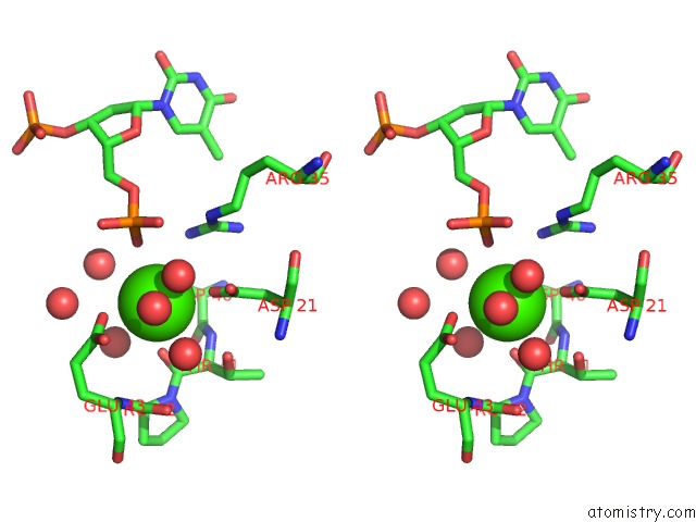
Stereo pair view
Mono view
Stereo pair view
A full contact list of Calcium with other atoms in the Ca binding
site number 1 of Crystal Structure of Staphylococcal Nuclease Variant Delta+Phs A90R at Cryogenic Temperature within 5.0Å range:
|
Reference:
M.J.Harms,
J.L.Schlessman,
G.R.Sue,
B.Garcia-Moreno E.
Arginine Residues at Internal Positions in A Protein Are Always Charged. Proc.Natl.Acad.Sci.Usa V. 108 18954 2011.
ISSN: ISSN 0027-8424
PubMed: 22080604
DOI: 10.1073/PNAS.1104808108
Page generated: Sat Jul 13 09:03:06 2024
ISSN: ISSN 0027-8424
PubMed: 22080604
DOI: 10.1073/PNAS.1104808108
Last articles
Zn in 9J0NZn in 9J0O
Zn in 9J0P
Zn in 9FJX
Zn in 9EKB
Zn in 9C0F
Zn in 9CAH
Zn in 9CH0
Zn in 9CH3
Zn in 9CH1