Calcium »
PDB 3dcq-3dsw »
3dnz »
Calcium in PDB 3dnz: Thermolysin By Lb Nanotemplate Method Before High X-Ray Dose on Esrf ID14-2 Beamline
Enzymatic activity of Thermolysin By Lb Nanotemplate Method Before High X-Ray Dose on Esrf ID14-2 Beamline
All present enzymatic activity of Thermolysin By Lb Nanotemplate Method Before High X-Ray Dose on Esrf ID14-2 Beamline:
3.4.24.27;
3.4.24.27;
Protein crystallography data
The structure of Thermolysin By Lb Nanotemplate Method Before High X-Ray Dose on Esrf ID14-2 Beamline, PDB code: 3dnz
was solved by
E.Pechkova,
S.K.Tripathi,
C.Nicolini,
with X-Ray Crystallography technique. A brief refinement statistics is given in the table below:
| Resolution Low / High (Å) | 68.04 / 1.20 |
| Space group | P 61 2 2 |
| Cell size a, b, c (Å), α, β, γ (°) | 92.535, 92.535, 128.628, 90.00, 90.00, 120.00 |
| R / Rfree (%) | 24 / 26.7 |
Other elements in 3dnz:
The structure of Thermolysin By Lb Nanotemplate Method Before High X-Ray Dose on Esrf ID14-2 Beamline also contains other interesting chemical elements:
| Zinc | (Zn) | 1 atom |
Calcium Binding Sites:
The binding sites of Calcium atom in the Thermolysin By Lb Nanotemplate Method Before High X-Ray Dose on Esrf ID14-2 Beamline
(pdb code 3dnz). This binding sites where shown within
5.0 Angstroms radius around Calcium atom.
In total 4 binding sites of Calcium where determined in the Thermolysin By Lb Nanotemplate Method Before High X-Ray Dose on Esrf ID14-2 Beamline, PDB code: 3dnz:
Jump to Calcium binding site number: 1; 2; 3; 4;
In total 4 binding sites of Calcium where determined in the Thermolysin By Lb Nanotemplate Method Before High X-Ray Dose on Esrf ID14-2 Beamline, PDB code: 3dnz:
Jump to Calcium binding site number: 1; 2; 3; 4;
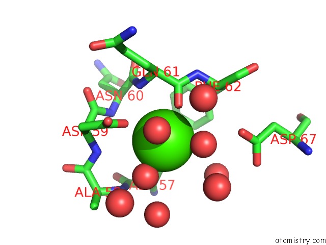
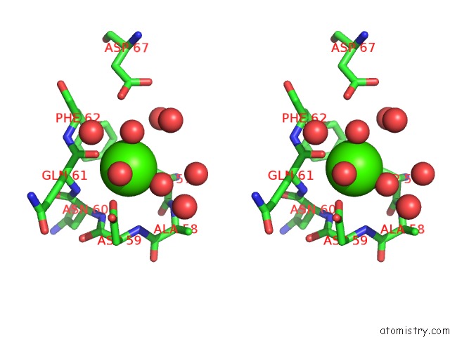
Calcium binding site 1 out of 4 in 3dnz
Go back to
Calcium binding site 1 out
of 4 in the Thermolysin By Lb Nanotemplate Method Before High X-Ray Dose on Esrf ID14-2 Beamline
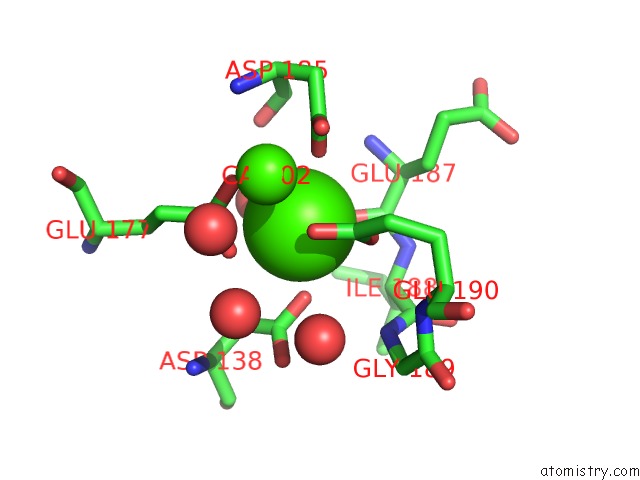
Mono view
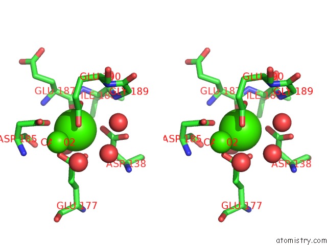
Stereo pair view
Mono view
Stereo pair view
A full contact list of Calcium with other atoms in the Ca binding
site number 1 of Thermolysin By Lb Nanotemplate Method Before High X-Ray Dose on Esrf ID14-2 Beamline within 5.0Å range:
|
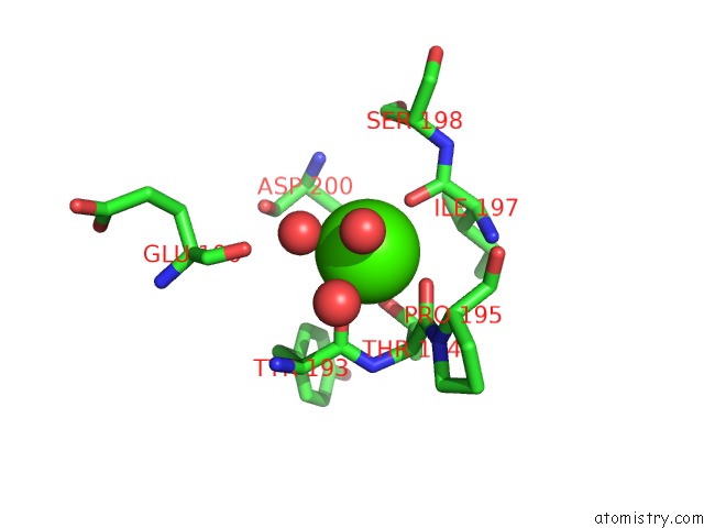
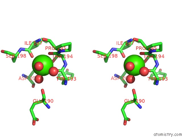
Calcium binding site 2 out of 4 in 3dnz
Go back to
Calcium binding site 2 out
of 4 in the Thermolysin By Lb Nanotemplate Method Before High X-Ray Dose on Esrf ID14-2 Beamline
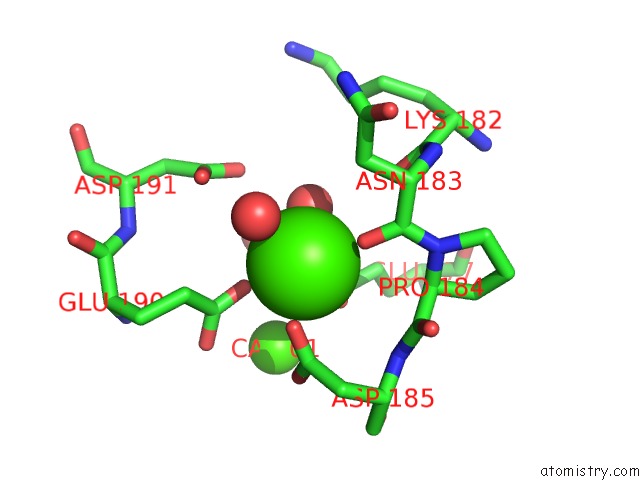
Mono view
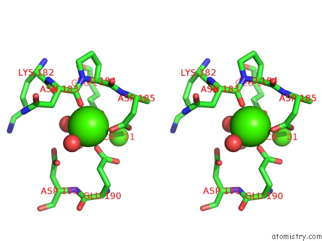
Stereo pair view
Mono view
Stereo pair view
A full contact list of Calcium with other atoms in the Ca binding
site number 2 of Thermolysin By Lb Nanotemplate Method Before High X-Ray Dose on Esrf ID14-2 Beamline within 5.0Å range:
|
Calcium binding site 3 out of 4 in 3dnz
Go back to
Calcium binding site 3 out
of 4 in the Thermolysin By Lb Nanotemplate Method Before High X-Ray Dose on Esrf ID14-2 Beamline
Mono view
Stereo pair view
Mono view
Stereo pair view
A full contact list of Calcium with other atoms in the Ca binding
site number 3 of Thermolysin By Lb Nanotemplate Method Before High X-Ray Dose on Esrf ID14-2 Beamline within 5.0Å range:
|
Calcium binding site 4 out of 4 in 3dnz
Go back to
Calcium binding site 4 out
of 4 in the Thermolysin By Lb Nanotemplate Method Before High X-Ray Dose on Esrf ID14-2 Beamline
Mono view
Stereo pair view
Mono view
Stereo pair view
A full contact list of Calcium with other atoms in the Ca binding
site number 4 of Thermolysin By Lb Nanotemplate Method Before High X-Ray Dose on Esrf ID14-2 Beamline within 5.0Å range:
|
Reference:
E.Pechkova,
S.K.Tripathi,
C.Nicolini.
Radiation Damage in Protein Structural Characterization By Synchrotron Radiation: State of the Art and Nanotechnology-Based Perspective To Be Published.
Page generated: Tue Jul 8 11:40:01 2025
Last articles
Cl in 8F7LCl in 8F6G
Cl in 8F5A
Cl in 8F5Y
Cl in 8F5J
Cl in 8F58
Cl in 8F55
Cl in 8F4Z
Cl in 8F57
Cl in 8F4K