Calcium »
PDB 3dcq-3dsw »
3do1 »
Calcium in PDB 3do1: Thermolysin By Classical Hanging Drop Method Before High X- Ray Dose on Esrf ID14-2 Beamline
Enzymatic activity of Thermolysin By Classical Hanging Drop Method Before High X- Ray Dose on Esrf ID14-2 Beamline
All present enzymatic activity of Thermolysin By Classical Hanging Drop Method Before High X- Ray Dose on Esrf ID14-2 Beamline:
3.4.24.27;
3.4.24.27;
Protein crystallography data
The structure of Thermolysin By Classical Hanging Drop Method Before High X- Ray Dose on Esrf ID14-2 Beamline, PDB code: 3do1
was solved by
E.Pechkova,
S.K.Tripathi,
C.Nicolini,
with X-Ray Crystallography technique. A brief refinement statistics is given in the table below:
| Resolution Low / High (Å) | 68.04 / 1.33 |
| Space group | P 61 2 2 |
| Cell size a, b, c (Å), α, β, γ (°) | 92.526, 92.526, 128.553, 90.00, 90.00, 120.00 |
| R / Rfree (%) | 23.1 / 27.7 |
Other elements in 3do1:
The structure of Thermolysin By Classical Hanging Drop Method Before High X- Ray Dose on Esrf ID14-2 Beamline also contains other interesting chemical elements:
| Zinc | (Zn) | 1 atom |
Calcium Binding Sites:
The binding sites of Calcium atom in the Thermolysin By Classical Hanging Drop Method Before High X- Ray Dose on Esrf ID14-2 Beamline
(pdb code 3do1). This binding sites where shown within
5.0 Angstroms radius around Calcium atom.
In total 4 binding sites of Calcium where determined in the Thermolysin By Classical Hanging Drop Method Before High X- Ray Dose on Esrf ID14-2 Beamline, PDB code: 3do1:
Jump to Calcium binding site number: 1; 2; 3; 4;
In total 4 binding sites of Calcium where determined in the Thermolysin By Classical Hanging Drop Method Before High X- Ray Dose on Esrf ID14-2 Beamline, PDB code: 3do1:
Jump to Calcium binding site number: 1; 2; 3; 4;
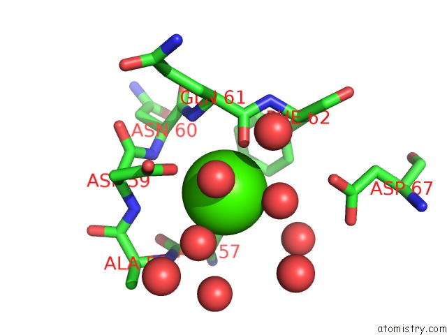
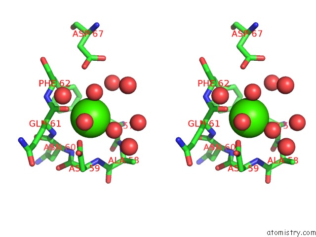
Calcium binding site 1 out of 4 in 3do1
Go back to
Calcium binding site 1 out
of 4 in the Thermolysin By Classical Hanging Drop Method Before High X- Ray Dose on Esrf ID14-2 Beamline
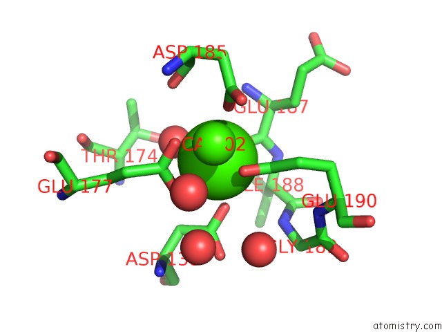
Mono view
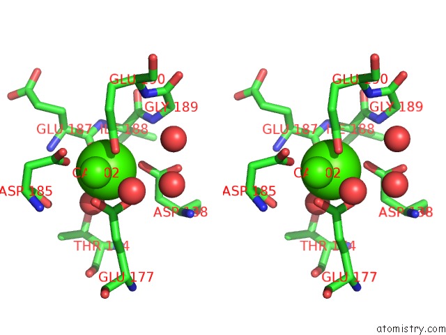
Stereo pair view
Mono view
Stereo pair view
A full contact list of Calcium with other atoms in the Ca binding
site number 1 of Thermolysin By Classical Hanging Drop Method Before High X- Ray Dose on Esrf ID14-2 Beamline within 5.0Å range:
|
Calcium binding site 2 out of 4 in 3do1
Go back to
Calcium binding site 2 out
of 4 in the Thermolysin By Classical Hanging Drop Method Before High X- Ray Dose on Esrf ID14-2 Beamline
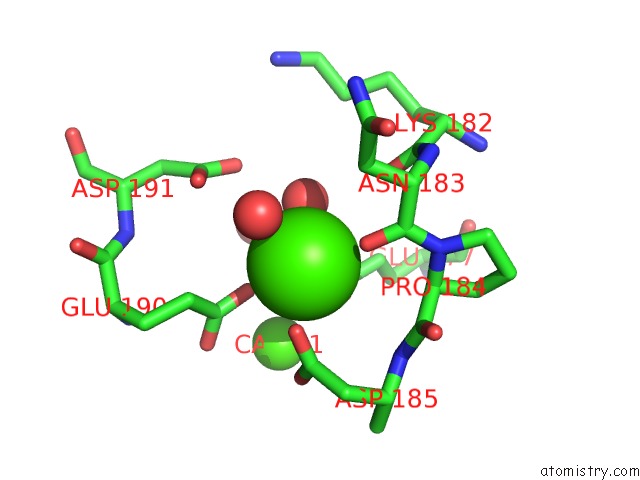
Mono view
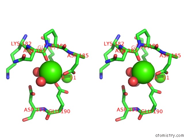
Stereo pair view
Mono view
Stereo pair view
A full contact list of Calcium with other atoms in the Ca binding
site number 2 of Thermolysin By Classical Hanging Drop Method Before High X- Ray Dose on Esrf ID14-2 Beamline within 5.0Å range:
|
Calcium binding site 3 out of 4 in 3do1
Go back to
Calcium binding site 3 out
of 4 in the Thermolysin By Classical Hanging Drop Method Before High X- Ray Dose on Esrf ID14-2 Beamline
Mono view
Stereo pair view
Mono view
Stereo pair view
A full contact list of Calcium with other atoms in the Ca binding
site number 3 of Thermolysin By Classical Hanging Drop Method Before High X- Ray Dose on Esrf ID14-2 Beamline within 5.0Å range:
|
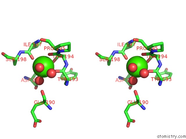
Calcium binding site 4 out of 4 in 3do1
Go back to
Calcium binding site 4 out
of 4 in the Thermolysin By Classical Hanging Drop Method Before High X- Ray Dose on Esrf ID14-2 Beamline

Mono view
Stereo pair view
Mono view
Stereo pair view
A full contact list of Calcium with other atoms in the Ca binding
site number 4 of Thermolysin By Classical Hanging Drop Method Before High X- Ray Dose on Esrf ID14-2 Beamline within 5.0Å range:
|
Reference:
E.Pechkova,
S.K.Tripathi,
C.Nicolini.
Radiation Damage in Protein Structural Characterization By Synchrotron Radiation: State of the Art and Nanotechnology-Based Perspective To Be Published.
Page generated: Sat Jul 13 09:04:59 2024
Last articles
Zn in 9J0NZn in 9J0O
Zn in 9J0P
Zn in 9FJX
Zn in 9EKB
Zn in 9C0F
Zn in 9CAH
Zn in 9CH0
Zn in 9CH3
Zn in 9CH1