Calcium »
PDB 3dsx-3ead »
3dtu »
Calcium in PDB 3dtu: Catalytic Core Subunits (I and II) of Cytochrome C Oxidase From Rhodobacter Sphaeroides Complexed with Deoxycholic Acid
Enzymatic activity of Catalytic Core Subunits (I and II) of Cytochrome C Oxidase From Rhodobacter Sphaeroides Complexed with Deoxycholic Acid
All present enzymatic activity of Catalytic Core Subunits (I and II) of Cytochrome C Oxidase From Rhodobacter Sphaeroides Complexed with Deoxycholic Acid:
1.9.3.1;
1.9.3.1;
Protein crystallography data
The structure of Catalytic Core Subunits (I and II) of Cytochrome C Oxidase From Rhodobacter Sphaeroides Complexed with Deoxycholic Acid, PDB code: 3dtu
was solved by
L.Qin,
D.A.Mills,
L.Buhrow,
C.Hiser,
S.Ferguson-Miller,
with X-Ray Crystallography technique. A brief refinement statistics is given in the table below:
| Resolution Low / High (Å) | 40.00 / 2.15 |
| Space group | P 21 21 21 |
| Cell size a, b, c (Å), α, β, γ (°) | 123.243, 132.052, 167.966, 90.00, 90.00, 90.00 |
| R / Rfree (%) | 18.4 / 21.2 |
Other elements in 3dtu:
The structure of Catalytic Core Subunits (I and II) of Cytochrome C Oxidase From Rhodobacter Sphaeroides Complexed with Deoxycholic Acid also contains other interesting chemical elements:
| Magnesium | (Mg) | 2 atoms |
| Cadmium | (Cd) | 5 atoms |
| Iron | (Fe) | 4 atoms |
| Copper | (Cu) | 6 atoms |
Calcium Binding Sites:
The binding sites of Calcium atom in the Catalytic Core Subunits (I and II) of Cytochrome C Oxidase From Rhodobacter Sphaeroides Complexed with Deoxycholic Acid
(pdb code 3dtu). This binding sites where shown within
5.0 Angstroms radius around Calcium atom.
In total 2 binding sites of Calcium where determined in the Catalytic Core Subunits (I and II) of Cytochrome C Oxidase From Rhodobacter Sphaeroides Complexed with Deoxycholic Acid, PDB code: 3dtu:
Jump to Calcium binding site number: 1; 2;
In total 2 binding sites of Calcium where determined in the Catalytic Core Subunits (I and II) of Cytochrome C Oxidase From Rhodobacter Sphaeroides Complexed with Deoxycholic Acid, PDB code: 3dtu:
Jump to Calcium binding site number: 1; 2;
Calcium binding site 1 out of 2 in 3dtu
Go back to
Calcium binding site 1 out
of 2 in the Catalytic Core Subunits (I and II) of Cytochrome C Oxidase From Rhodobacter Sphaeroides Complexed with Deoxycholic Acid
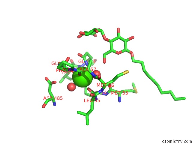
Mono view
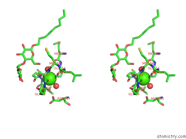
Stereo pair view
Mono view
Stereo pair view
A full contact list of Calcium with other atoms in the Ca binding
site number 1 of Catalytic Core Subunits (I and II) of Cytochrome C Oxidase From Rhodobacter Sphaeroides Complexed with Deoxycholic Acid within 5.0Å range:
|
Calcium binding site 2 out of 2 in 3dtu
Go back to
Calcium binding site 2 out
of 2 in the Catalytic Core Subunits (I and II) of Cytochrome C Oxidase From Rhodobacter Sphaeroides Complexed with Deoxycholic Acid
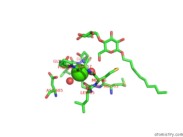
Mono view
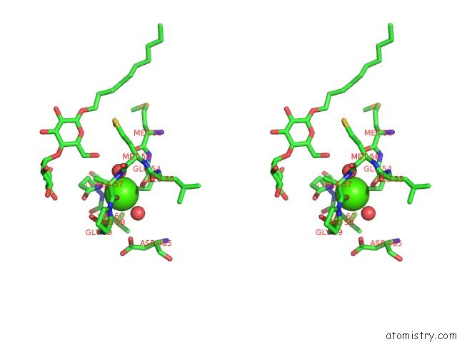
Stereo pair view
Mono view
Stereo pair view
A full contact list of Calcium with other atoms in the Ca binding
site number 2 of Catalytic Core Subunits (I and II) of Cytochrome C Oxidase From Rhodobacter Sphaeroides Complexed with Deoxycholic Acid within 5.0Å range:
|
Reference:
L.Qin,
D.A.Mills,
L.Buhrow,
C.Hiser,
S.Ferguson-Miller.
A Conserved Steroid Binding Site in Cytochrome C Oxidase. Biochemistry V. 47 9931 2008.
ISSN: ISSN 0006-2960
PubMed: 18759498
DOI: 10.1021/BI8013483
Page generated: Tue Jul 8 11:43:32 2025
ISSN: ISSN 0006-2960
PubMed: 18759498
DOI: 10.1021/BI8013483
Last articles
Cl in 8AXRCl in 8AWW
Cl in 8AXE
Cl in 8AUT
Cl in 8AWN
Cl in 8AW2
Cl in 8AWG
Cl in 8AV7
Cl in 8AV9
Cl in 8AV5