Calcium »
PDB 3dsx-3ead »
3e4q »
Calcium in PDB 3e4q: Crystal Structure of Apo Dctb
Enzymatic activity of Crystal Structure of Apo Dctb
All present enzymatic activity of Crystal Structure of Apo Dctb:
2.7.13.3;
2.7.13.3;
Protein crystallography data
The structure of Crystal Structure of Apo Dctb, PDB code: 3e4q
was solved by
Y.F.Zhou,
J.Nan,
B.Y.Nan,
Y.H.Liang,
S.Panjikar,
X.D.Su,
with X-Ray Crystallography technique. A brief refinement statistics is given in the table below:
| Resolution Low / High (Å) | 30.00 / 2.75 |
| Space group | P 1 21 1 |
| Cell size a, b, c (Å), α, β, γ (°) | 57.988, 38.735, 111.172, 90.00, 94.47, 90.00 |
| R / Rfree (%) | 19.5 / 27.4 |
Calcium Binding Sites:
The binding sites of Calcium atom in the Crystal Structure of Apo Dctb
(pdb code 3e4q). This binding sites where shown within
5.0 Angstroms radius around Calcium atom.
In total 2 binding sites of Calcium where determined in the Crystal Structure of Apo Dctb, PDB code: 3e4q:
Jump to Calcium binding site number: 1; 2;
In total 2 binding sites of Calcium where determined in the Crystal Structure of Apo Dctb, PDB code: 3e4q:
Jump to Calcium binding site number: 1; 2;
Calcium binding site 1 out of 2 in 3e4q
Go back to
Calcium binding site 1 out
of 2 in the Crystal Structure of Apo Dctb
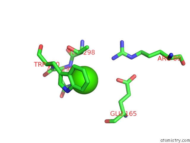
Mono view
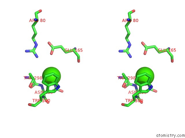
Stereo pair view
Mono view
Stereo pair view
A full contact list of Calcium with other atoms in the Ca binding
site number 1 of Crystal Structure of Apo Dctb within 5.0Å range:
|
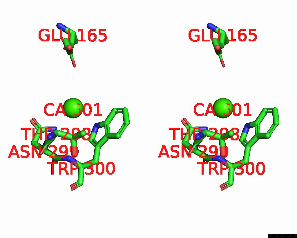
Calcium binding site 2 out of 2 in 3e4q
Go back to
Calcium binding site 2 out
of 2 in the Crystal Structure of Apo Dctb

Mono view
Stereo pair view
Mono view
Stereo pair view
A full contact list of Calcium with other atoms in the Ca binding
site number 2 of Crystal Structure of Apo Dctb within 5.0Å range:
|
Reference:
Y.F.Zhou,
B.Y.Nan,
J.Nan,
Q.J.Ma,
S.Panjikar,
Y.H.Liang,
Y.P.Wang,
X.D.Su.
C4-Dicarboxylates Sensing Mechanism Revealed By the Crystal Structures of Dctb Sensor Domain. J.Mol.Biol. V. 383 49 2008.
ISSN: ISSN 0022-2836
PubMed: 18725229
DOI: 10.1016/J.JMB.2008.08.010
Page generated: Tue Jul 8 11:48:16 2025
ISSN: ISSN 0022-2836
PubMed: 18725229
DOI: 10.1016/J.JMB.2008.08.010
Last articles
Cl in 8CE2Cl in 8CCR
Cl in 8CCM
Cl in 8CCF
Cl in 8CCE
Cl in 8CCB
Cl in 8CCD
Cl in 8CCC
Cl in 8CC6
Cl in 8CBH