Calcium »
PDB 3ecj-3eqd »
3edy »
Calcium in PDB 3edy: Crystal Structure of the Precursor Form of Human Tripeptidyl-Peptidase 1
Enzymatic activity of Crystal Structure of the Precursor Form of Human Tripeptidyl-Peptidase 1
All present enzymatic activity of Crystal Structure of the Precursor Form of Human Tripeptidyl-Peptidase 1:
3.4.14.9;
3.4.14.9;
Protein crystallography data
The structure of Crystal Structure of the Precursor Form of Human Tripeptidyl-Peptidase 1, PDB code: 3edy
was solved by
J.Guhaniyogi,
I.Sohar,
K.Das,
P.Lobel,
A.M.Stock,
with X-Ray Crystallography technique. A brief refinement statistics is given in the table below:
| Resolution Low / High (Å) | 29.72 / 1.85 |
| Space group | P 21 21 21 |
| Cell size a, b, c (Å), α, β, γ (°) | 59.807, 93.173, 102.479, 90.00, 90.00, 90.00 |
| R / Rfree (%) | 17.9 / 20.5 |
Calcium Binding Sites:
The binding sites of Calcium atom in the Crystal Structure of the Precursor Form of Human Tripeptidyl-Peptidase 1
(pdb code 3edy). This binding sites where shown within
5.0 Angstroms radius around Calcium atom.
In total only one binding site of Calcium was determined in the Crystal Structure of the Precursor Form of Human Tripeptidyl-Peptidase 1, PDB code: 3edy:
In total only one binding site of Calcium was determined in the Crystal Structure of the Precursor Form of Human Tripeptidyl-Peptidase 1, PDB code: 3edy:
Calcium binding site 1 out of 1 in 3edy
Go back to
Calcium binding site 1 out
of 1 in the Crystal Structure of the Precursor Form of Human Tripeptidyl-Peptidase 1
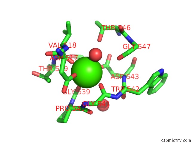
Mono view
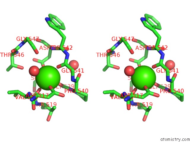
Stereo pair view
Mono view
Stereo pair view
A full contact list of Calcium with other atoms in the Ca binding
site number 1 of Crystal Structure of the Precursor Form of Human Tripeptidyl-Peptidase 1 within 5.0Å range:
|
Reference:
J.Guhaniyogi,
I.Sohar,
K.Das,
A.M.Stock,
P.Lobel.
Crystal Structure and Autoactivation Pathway of the Precursor Form of Human Tripeptidyl-Peptidase 1, the Enzyme Deficient in Late Infantile Ceroid Lipofuscinosis J.Biol.Chem. V. 284 3985 2009.
ISSN: ISSN 0021-9258
PubMed: 19038967
DOI: 10.1074/JBC.M806943200
Page generated: Sat Jul 13 09:23:02 2024
ISSN: ISSN 0021-9258
PubMed: 19038967
DOI: 10.1074/JBC.M806943200
Last articles
Zn in 9J0NZn in 9J0O
Zn in 9J0P
Zn in 9FJX
Zn in 9EKB
Zn in 9C0F
Zn in 9CAH
Zn in 9CH0
Zn in 9CH3
Zn in 9CH1