Calcium »
PDB 3ecj-3eqd »
3ee6 »
Calcium in PDB 3ee6: Crystal Structure Analysis of Tripeptidyl Peptidase -I
Enzymatic activity of Crystal Structure Analysis of Tripeptidyl Peptidase -I
All present enzymatic activity of Crystal Structure Analysis of Tripeptidyl Peptidase -I:
3.4.14.9;
3.4.14.9;
Protein crystallography data
The structure of Crystal Structure Analysis of Tripeptidyl Peptidase -I, PDB code: 3ee6
was solved by
A.Pal,
R.Kraetzner,
M.Grapp,
T.Gruene,
K.Schreiber,
M.Granborg,
H.Urlaub,
A.R.Asif,
S.Becker,
J.Gartner,
G.M.Sheldrick,
R.Steinfeld,
with X-Ray Crystallography technique. A brief refinement statistics is given in the table below:
| Resolution Low / High (Å) | 39.21 / 2.35 |
| Space group | P 21 21 2 |
| Cell size a, b, c (Å), α, β, γ (°) | 113.450, 128.930, 100.500, 90.00, 90.00, 90.00 |
| R / Rfree (%) | 21.8 / 26.2 |
Other elements in 3ee6:
The structure of Crystal Structure Analysis of Tripeptidyl Peptidase -I also contains other interesting chemical elements:
| Chlorine | (Cl) | 2 atoms |
| Zinc | (Zn) | 8 atoms |
Calcium Binding Sites:
The binding sites of Calcium atom in the Crystal Structure Analysis of Tripeptidyl Peptidase -I
(pdb code 3ee6). This binding sites where shown within
5.0 Angstroms radius around Calcium atom.
In total 2 binding sites of Calcium where determined in the Crystal Structure Analysis of Tripeptidyl Peptidase -I, PDB code: 3ee6:
Jump to Calcium binding site number: 1; 2;
In total 2 binding sites of Calcium where determined in the Crystal Structure Analysis of Tripeptidyl Peptidase -I, PDB code: 3ee6:
Jump to Calcium binding site number: 1; 2;
Calcium binding site 1 out of 2 in 3ee6
Go back to
Calcium binding site 1 out
of 2 in the Crystal Structure Analysis of Tripeptidyl Peptidase -I
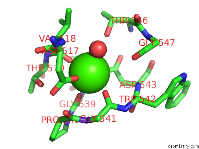
Mono view
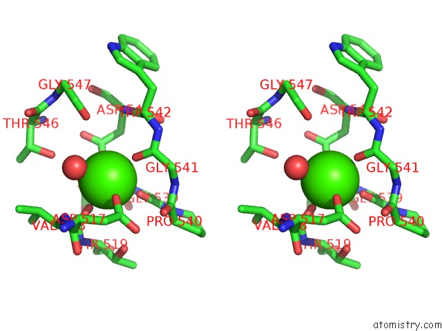
Stereo pair view
Mono view
Stereo pair view
A full contact list of Calcium with other atoms in the Ca binding
site number 1 of Crystal Structure Analysis of Tripeptidyl Peptidase -I within 5.0Å range:
|
Calcium binding site 2 out of 2 in 3ee6
Go back to
Calcium binding site 2 out
of 2 in the Crystal Structure Analysis of Tripeptidyl Peptidase -I
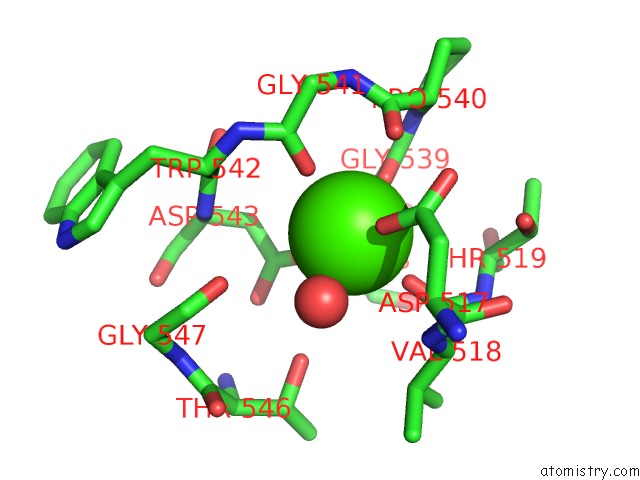
Mono view
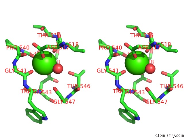
Stereo pair view
Mono view
Stereo pair view
A full contact list of Calcium with other atoms in the Ca binding
site number 2 of Crystal Structure Analysis of Tripeptidyl Peptidase -I within 5.0Å range:
|
Reference:
A.Pal,
R.Kraetzner,
T.Gruene,
M.Grapp,
K.Schreiber,
M.Gronborg,
H.Urlaub,
S.Becker,
A.R.Asif,
J.Gartner,
G.M.Sheldrick,
R.Steinfeld.
Structure of Tripeptidyl-Peptidase I Provides Insight Into the Molecular Basis of Late Infantile Neuronal Ceroid Lipofuscinosis J.Biol.Chem. V. 284 3976 2009.
ISSN: ISSN 0021-9258
PubMed: 19038966
DOI: 10.1074/JBC.M806947200
Page generated: Tue Jul 8 11:52:38 2025
ISSN: ISSN 0021-9258
PubMed: 19038966
DOI: 10.1074/JBC.M806947200
Last articles
F in 7M0YF in 7M0Z
F in 7M0M
F in 7M0V
F in 7M0W
F in 7M0U
F in 7M0N
F in 7M0T
F in 7M00
F in 7M01